
Homework Answers
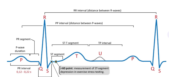
A) P wavve
B) QRS wave
C) ST segment
D) T wave
Please do rate and liked me...........
Add Answer to:
Label segments of the ECG
Use the following terms to label the the ECG Diagram. -...
Procedure 1 Interpreting an ECG You will now perform some basic ECG interpretation. Following are two...
 Procedure 1 Interpreting an ECG You will now perform some basic ECG interpretation. Following are two tracings for which you will calculate the heart duration of key periods of the ECG. 1 Identify and label the P wave, QRS complex, T wave, P-R interval, R-R interval, and Q-T interval on Tracings 1 and 2 in Figure 11.27 Tracing 1 Tracing 2 FIGURE 11.27 ECG tracings 2 Cakulate the heart rate for each tracing, Are the values normal or abnormal? Heart...
match the part of the ecg with the correct description + R R Q S P-R...
 match the part of the ecg with the correct description
+ R R Q S P-R segment S-T segment Millivolts P wave T wave PR interval QT interval QRS complex P Wave PR interval QRS Complex A. The sinoatrial node fires, depolarizing the atria The entire period in which ventricles depolarize, then B. repolarize. Represents the complete duration of ventricular systole Electrical signal causing depolarization travels from the sinoatrial node to the atrioventricular node D. The right and left ventricles...
match the part of the ecg with the correct description
+ R R Q S P-R segment S-T segment Millivolts P wave T wave PR interval QT interval QRS complex P Wave PR interval QRS Complex A. The sinoatrial node fires, depolarizing the atria The entire period in which ventricles depolarize, then B. repolarize. Represents the complete duration of ventricular systole Electrical signal causing depolarization travels from the sinoatrial node to the atrioventricular node D. The right and left ventricles...
i dont under stand how to figure out the bpm with this information Search... Heart Physiology ECG and nd ea Cegle Br...
 i dont under stand how to figure out the bpm with this
information
Search... Heart Physiology ECG and nd ea Cegle Brightspace aMyCampus G Google (2) Lab Notebook ECG Components Heart Rate bpm ECG Component Cycle 1 Cycle 2 Cycle 3 Mean Normal Duration P wave 11 11 12 0.11s 0.06-0.11 PR Interval 22 0.23s 0.12-0.20 23 24 PR segment 17 13 0.14s 0.08 12 QRS complex 08 07 08 0.08s <0.12 0.34s 0.36-0.44 33 QT interval 38 31 ST...
i dont under stand how to figure out the bpm with this
information
Search... Heart Physiology ECG and nd ea Cegle Brightspace aMyCampus G Google (2) Lab Notebook ECG Components Heart Rate bpm ECG Component Cycle 1 Cycle 2 Cycle 3 Mean Normal Duration P wave 11 11 12 0.11s 0.06-0.11 PR Interval 22 0.23s 0.12-0.20 23 24 PR segment 17 13 0.14s 0.08 12 QRS complex 08 07 08 0.08s <0.12 0.34s 0.36-0.44 33 QT interval 38 31 ST...
Procedure 1 Correlating Points on an ECG with Electrical Events of the Heart Let's start by...
 Procedure 1 Correlating Points on an ECG with Electrical Events of the Heart Let's start by correlating the following points on the ECG with the electrical activity occurring in the heart Identify the electrical events taking place in cardiac cells at each of the six points int the ECG in Figure 19.8, and explain the events in the answer lines provided. Do remember though that the ECG shows only electrical events, not mechanical events. A heart can have electrical activity...
QUESTION 21 A farmer standing in a field of oats in Scotland (in other words, not...
QUESTION 21 A farmer standing in a field of oats in Scotland (in other words, not in high mountains) is most likely to experience which form of lightning strike? a) A ground current b) A side-strike c) A direct strike 1 points QUESTION 22 What do we see on the ECG as the atria are getting ready to contract? a) The P wave b) The QRS wave c) The T wave 1 points QUESTION 23 During what part...
Compare the supine ECG values to the post-exercise values. Which intervals and/or segments changed most significantly?...
Compare the supine ECG values to the post-exercise values. Which intervals and/or segments changed most significantly? Include a detailed discussion of specific changes (include numerical values) to support your findings and analysis. Discuss your group’s heart rate data and use your knowledge of physiology to explain how and why these changes were seen. Address your hypothesis and explain the overall significance of the changes seen in cardiac cycle. Activity P-R Q-T QRS complex T-P Heart rate Supine (resting) 0.0770 0.350...
61. What state creates electrical activity that can be picked up on the ECG tracing? a...
 61. What state creates electrical activity that can be picked up on the ECG tracing? a Depolarized state b. Polarized state c. Repolarized state d. All of the above e. A and C Upward movement away from the baseline in an ECG tracing. a. Isoelectrie line b. Negative deflection c. Positive deflection d. Complex e. Wave 63. A period of time between two points or events on the ECG tracing a. Wave b. Complex c. Isoelectric line d. Segment e....
Fill out the heart rate table in the sitting tab Lab Notebook Standing Sitting After Exercise...
 Fill out the heart rate table in the sitting tab
Lab Notebook Standing Sitting After Exercise Cycle 2 ECG Component Cycle 1 Cycle 3 Mean Normal Duration Os 0.06-0.11 P wave P-R interval P-R segment Os 0.12-0.20 Os 0.08 Os <0.12 Os 0.36-0.44 QRS complex Q-T interval ST segment Os 0.12 Os 0.16 T wave Amplitude Omv <0.25 Stand P wave
Fill out the heart rate table in the sitting tab
Lab Notebook Standing Sitting After Exercise Cycle 2 ECG Component Cycle 1 Cycle 3 Mean Normal Duration Os 0.06-0.11 P wave P-R interval P-R segment Os 0.12-0.20 Os 0.08 Os <0.12 Os 0.36-0.44 QRS complex Q-T interval ST segment Os 0.12 Os 0.16 T wave Amplitude Omv <0.25 Stand P wave
Fill out the heart rate table in the sitting tab Lab Notebook Standing Sitting After Exercise...
 Fill out the heart rate table in the sitting tab
Lab Notebook Standing Sitting After Exercise Cycle 2 ECG Component Cycle 1 Cycle 3 Mean Normal Duration Os 0.06-0.11 P wave P-R interval P-R segment Os 0.12-0.20 Os 0.08 Os <0.12 Os 0.36-0.44 QRS complex Q-T interval ST segment Os 0.12 Os 0.16 T wave Amplitude Omv <0.25 Stand P wave
Fill out the heart rate table in the sitting tab
Lab Notebook Standing Sitting After Exercise Cycle 2 ECG Component Cycle 1 Cycle 3 Mean Normal Duration Os 0.06-0.11 P wave P-R interval P-R segment Os 0.12-0.20 Os 0.08 Os <0.12 Os 0.36-0.44 QRS complex Q-T interval ST segment Os 0.12 Os 0.16 T wave Amplitude Omv <0.25 Stand P wave
Identify the electrical activity associated with each of the ECG components and indicate whether the electrical...
Identify the electrical activity associated with each of the ECG components and indicate whether the electrical activity in the heart has the same or opposite polarity as Lead II. • P wave: • QRS complex: • T wave:
 Procedure 1 Interpreting an ECG You will now perform some basic ECG interpretation. Following are two tracings for which you will calculate the heart duration of key periods of the ECG. 1 Identify and label the P wave, QRS complex, T wave, P-R interval, R-R interval, and Q-T interval on Tracings 1 and 2 in Figure 11.27 Tracing 1 Tracing 2 FIGURE 11.27 ECG tracings 2 Cakulate the heart rate for each tracing, Are the values normal or abnormal? Heart...
 match the part of the ecg with the correct description
+ R R Q S P-R segment S-T segment Millivolts P wave T wave PR interval QT interval QRS complex P Wave PR interval QRS Complex A. The sinoatrial node fires, depolarizing the atria The entire period in which ventricles depolarize, then B. repolarize. Represents the complete duration of ventricular systole Electrical signal causing depolarization travels from the sinoatrial node to the atrioventricular node D. The right and left ventricles...
match the part of the ecg with the correct description
+ R R Q S P-R segment S-T segment Millivolts P wave T wave PR interval QT interval QRS complex P Wave PR interval QRS Complex A. The sinoatrial node fires, depolarizing the atria The entire period in which ventricles depolarize, then B. repolarize. Represents the complete duration of ventricular systole Electrical signal causing depolarization travels from the sinoatrial node to the atrioventricular node D. The right and left ventricles...
 i dont under stand how to figure out the bpm with this
information
Search... Heart Physiology ECG and nd ea Cegle Brightspace aMyCampus G Google (2) Lab Notebook ECG Components Heart Rate bpm ECG Component Cycle 1 Cycle 2 Cycle 3 Mean Normal Duration P wave 11 11 12 0.11s 0.06-0.11 PR Interval 22 0.23s 0.12-0.20 23 24 PR segment 17 13 0.14s 0.08 12 QRS complex 08 07 08 0.08s <0.12 0.34s 0.36-0.44 33 QT interval 38 31 ST...
i dont under stand how to figure out the bpm with this
information
Search... Heart Physiology ECG and nd ea Cegle Brightspace aMyCampus G Google (2) Lab Notebook ECG Components Heart Rate bpm ECG Component Cycle 1 Cycle 2 Cycle 3 Mean Normal Duration P wave 11 11 12 0.11s 0.06-0.11 PR Interval 22 0.23s 0.12-0.20 23 24 PR segment 17 13 0.14s 0.08 12 QRS complex 08 07 08 0.08s <0.12 0.34s 0.36-0.44 33 QT interval 38 31 ST...
 Procedure 1 Correlating Points on an ECG with Electrical Events of the Heart Let's start by correlating the following points on the ECG with the electrical activity occurring in the heart Identify the electrical events taking place in cardiac cells at each of the six points int the ECG in Figure 19.8, and explain the events in the answer lines provided. Do remember though that the ECG shows only electrical events, not mechanical events. A heart can have electrical activity...
 61. What state creates electrical activity that can be picked up on the ECG tracing? a Depolarized state b. Polarized state c. Repolarized state d. All of the above e. A and C Upward movement away from the baseline in an ECG tracing. a. Isoelectrie line b. Negative deflection c. Positive deflection d. Complex e. Wave 63. A period of time between two points or events on the ECG tracing a. Wave b. Complex c. Isoelectric line d. Segment e....
 Fill out the heart rate table in the sitting tab
Lab Notebook Standing Sitting After Exercise Cycle 2 ECG Component Cycle 1 Cycle 3 Mean Normal Duration Os 0.06-0.11 P wave P-R interval P-R segment Os 0.12-0.20 Os 0.08 Os <0.12 Os 0.36-0.44 QRS complex Q-T interval ST segment Os 0.12 Os 0.16 T wave Amplitude Omv <0.25 Stand P wave
Fill out the heart rate table in the sitting tab
Lab Notebook Standing Sitting After Exercise Cycle 2 ECG Component Cycle 1 Cycle 3 Mean Normal Duration Os 0.06-0.11 P wave P-R interval P-R segment Os 0.12-0.20 Os 0.08 Os <0.12 Os 0.36-0.44 QRS complex Q-T interval ST segment Os 0.12 Os 0.16 T wave Amplitude Omv <0.25 Stand P wave
 Fill out the heart rate table in the sitting tab
Lab Notebook Standing Sitting After Exercise Cycle 2 ECG Component Cycle 1 Cycle 3 Mean Normal Duration Os 0.06-0.11 P wave P-R interval P-R segment Os 0.12-0.20 Os 0.08 Os <0.12 Os 0.36-0.44 QRS complex Q-T interval ST segment Os 0.12 Os 0.16 T wave Amplitude Omv <0.25 Stand P wave
Fill out the heart rate table in the sitting tab
Lab Notebook Standing Sitting After Exercise Cycle 2 ECG Component Cycle 1 Cycle 3 Mean Normal Duration Os 0.06-0.11 P wave P-R interval P-R segment Os 0.12-0.20 Os 0.08 Os <0.12 Os 0.36-0.44 QRS complex Q-T interval ST segment Os 0.12 Os 0.16 T wave Amplitude Omv <0.25 Stand P wave
Most questions answered within 3 hours.
-
Where is the error in this code sequence?
String s1 = "Hello";
String s2 = "ello";...
asked 11 months ago -
Financial data for Joel de Paris, Inc., for last year
follow:
Joel de Paris, Inc.
Balance...
asked 11 months ago -
Consider this reaction:
Al2(SO4)3 (aq)+ BaCl3
(aq) Al2Cl6 (aq)- +
3BaSO4(s) . What is the...
asked 11 months ago -
Suppose that Savneet is considering increasing her
recent random sample from 20 car rentals to 40...
asked 11 months ago -
Trucks arrive at an unloading terminal at an average rate of 120
per hour.
Trucks arrive...
asked 11 months ago -
Why are methanol and ethanol completely soluble in water while
octanol is not very little soluble....
asked 11 months ago -
A facilities manager at a university reads in a research report
that the mean amount of...
asked 11 months ago -
When the CuSO4 is rehydrated by adding water to the anhydrous
compound, is this an endothermic...
asked 11 months ago -
A ray of sunlight is passing from diamond into crown glass; the
angle of incidence is...
asked 11 months ago -
A block of mass 0.249 kg is placed on top of a light, vertical
spring of...
asked 11 months ago -
how do the kidneys compensate in the presences of acidosis
a) trigger hyperventilate
b) reserve acid...
asked 11 months ago -
Question 501 pts
The rental rate of capital to the firm increases. Which of the
following...
asked 11 months ago