
Homework Answers


Add Answer to:
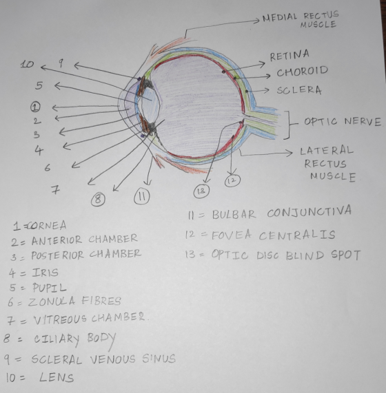
Pre-Lab Exercise 15-2 Anatomy of the Eye Label and color the structures of the eye in...
label all the parts of the ear please Pre-Lab Exercise 15-4 Anatomy of the Ear Label...
 label all the parts of the ear please
Pre-Lab Exercise 15-4 Anatomy of the Ear Label and color the structures of the car in Figure 15.4 with the terms from Exercise 15-2 (p. 403). Pob PESSO 000 009 sis
label all the parts of the ear please
Pre-Lab Exercise 15-4 Anatomy of the Ear Label and color the structures of the car in Figure 15.4 with the terms from Exercise 15-2 (p. 403). Pob PESSO 000 009 sis
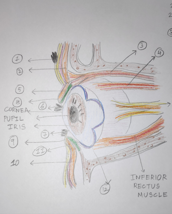
LAB 13 EXERCISE 13-7 EYE ANATOMY Label the following: Levator palpebrne m.. Tarsal plate, Lacrimal gland,...
 LAB 13 EXERCISE 13-7 EYE ANATOMY Label the following: Levator palpebrne m.. Tarsal plate, Lacrimal gland, Lacrimal ducts, Lacrimal Sac, Lacrimal caruncle, Medial canthus, Lateral canthus,
pre-lab exercise 21-1 Name Section Date PRE-LAB EXERCISES Complete the following esxercises prior to coming to...
 pre-lab exercise 21-1
Name Section Date PRE-LAB EXERCISES Complete the following esxercises prior to coming to lab, using your textbook and lab manvual for seference Pre-Lab Exercise 21-1 Key Terms You should be familiar with the following terms before coming to lab. Term General Structures of the Respiratory System Respiratory tract finition Parietal pleura Visceral pleura Pleural cavity Lungs and lobes Structures of the Respiratory Tract Nasal cavity Pharynx 21 Larynx Trachea Primary bronchi Secondary bronchi 510 I Exploring Anatomy...
pre-lab exercise 21-1
Name Section Date PRE-LAB EXERCISES Complete the following esxercises prior to coming to lab, using your textbook and lab manvual for seference Pre-Lab Exercise 21-1 Key Terms You should be familiar with the following terms before coming to lab. Term General Structures of the Respiratory System Respiratory tract finition Parietal pleura Visceral pleura Pleural cavity Lungs and lobes Structures of the Respiratory Tract Nasal cavity Pharynx 21 Larynx Trachea Primary bronchi Secondary bronchi 510 I Exploring Anatomy...
Using the figures provided, label the numbered structures of the cat's thoracic cavity 5. Using your...
 Using the figures provided, label
the numbered structures of the cat's thoracic cavity
5. Using your dissected specimen and Fig structures on Figure 20.4. cted specimen and Figures 20.2 and 20.3 as references, label the numbered (organ) (organ) - 15 forgan) Figure 20.4. Structures of the thoracic cavity. ventral view.photo to be labeled by students. Cervical Region 6. Using Figure 20.5 as a reference, locate the following structures: • trachea . parotid gland • mandibular gland • thyroid gland •...
Using the figures provided, label
the numbered structures of the cat's thoracic cavity
5. Using your dissected specimen and Fig structures on Figure 20.4. cted specimen and Figures 20.2 and 20.3 as references, label the numbered (organ) (organ) - 15 forgan) Figure 20.4. Structures of the thoracic cavity. ventral view.photo to be labeled by students. Cervical Region 6. Using Figure 20.5 as a reference, locate the following structures: • trachea . parotid gland • mandibular gland • thyroid gland •...
Pre-Lab #4 (This will help you prepare for the Skeletal Anatomy Test at end of Week...
Pre-Lab #4 (This will help you prepare for the Skeletal Anatomy Test at end of Week 5). Diagram (or obtain an unlabeled digital image of) and label the bones below. You can obtain unlabeled images online. For each bone complete the following tasks for Pre-Lab #4 Identify the bone and whether it is right or left (if that applies). Identify any markings or structures listed below on the bone (ex: fossa, cavity, process, etc). Identify any specific joints/articulations this bone...
Exercise 15.1: Gross Anatomy of the Ovaries and Uterine Tube 1. Review the location and gross...
 Exercise 15.1: Gross Anatomy of the Ovaries and Uterine Tube 1. Review the location and gross anatomical features of the ovaries and uterine tubes by studying models in the lab. Identify the following: a. Ovary f. Suspensory ligament k. Ampulla b. Cortex of ovary 9. Ovarian ligament I. Isthmus c. Medulla of ovary h. Uterine tube m. Mesosalpinx d. Mesovarium i. Infundibulum n. Mesometrium e. Broad ligament (2 parts) j. Fimbriae o. Cervix 2. Complete the labelling of Figure 15.2....
Anatomy and Physiology lab 568 Chapter Twenty-Four The Brain and Cranial Nerves Exercise 24.9 Teating Specific...
 Anatomy and Physiology lab 568 Chapter Twenty-Four The Brain and Cranial Nerves Exercise 24.9 Teating Specific Punetions of the Crenial Nerves 13. Match the disorder listed in column A with the cranial nerve associated with that disorder Isted in column B. Some answers may be used more than once. Column A 1 blindness 2 comeal reflex is absent (2 answers 3. difficuity turning the eye inferlor and lateral 4 inability to lateraly rotate the eye 5. Inability to maintain balance...
Instructors may of the Review SH using Masterin REVIEW SHEET Anatomy of the Respiratory System EXERCISE...
 Instructors may of the Review SH using Masterin REVIEW SHEET Anatomy of the Respiratory System EXERCISE Name Lab Time/Date Upper and lower Respiratory System Structures 1. Complete the labeling of the model of the respiratory structures (sagittal section) shown below. 2. Two pairs of mucosal folds are found in the larynx. Which pair are the true vocal cords (superior or infer 3. Name the specific cartilages in the larynx that correspond to the following descriptions. shaped like a ring forms...
Answer all the questions please LAB EXERCISE Name Primates Through the Miocene 1...
 Answer all the questions please
LAB EXERCISE Name Primates Through the Miocene 12.3 Section Date Materials Needed Station 1: Paleocene-The First Possible Primates (Alternative to Specimens: Figures A and B on page 336) 1 Compare a plesiadapiform primate with a strepsirhine. Fill in the table below with brief descriptions of the listed features for each. If using the figures provided rather than actual specimens, some features may not be visible. Station 1: A lemur or other strepsirhine anda Plesiadipis or...
Answer all the questions please
LAB EXERCISE Name Primates Through the Miocene 12.3 Section Date Materials Needed Station 1: Paleocene-The First Possible Primates (Alternative to Specimens: Figures A and B on page 336) 1 Compare a plesiadapiform primate with a strepsirhine. Fill in the table below with brief descriptions of the listed features for each. If using the figures provided rather than actual specimens, some features may not be visible. Station 1: A lemur or other strepsirhine anda Plesiadipis or...
Anatomy of the Heart 389 Right- ventricle - att ventricle Interventricular septum Entrance of inferior vena...
 Anatomy of the Heart 389 Right- ventricle - att ventricle Interventricular septum Entrance of inferior vena cava Cu surface ol wall pt night ventricie Fossa ovalis Peg in opening of coronary sinus Cusp of pulmonary valve Chordae tendineae Papillary Cusp of tricuspid valve muscle Wall of right ventricle (reflected) Moderator band Heart apex Figure 23.7 Right side of the sheep heart opened and reflected to reveal internal structures. Overview diagram illustrates the anatomical differences between the right and left ventricles....
 label all the parts of the ear please
Pre-Lab Exercise 15-4 Anatomy of the Ear Label and color the structures of the car in Figure 15.4 with the terms from Exercise 15-2 (p. 403). Pob PESSO 000 009 sis
label all the parts of the ear please
Pre-Lab Exercise 15-4 Anatomy of the Ear Label and color the structures of the car in Figure 15.4 with the terms from Exercise 15-2 (p. 403). Pob PESSO 000 009 sis
 LAB 13 EXERCISE 13-7 EYE ANATOMY Label the following: Levator palpebrne m.. Tarsal plate, Lacrimal gland, Lacrimal ducts, Lacrimal Sac, Lacrimal caruncle, Medial canthus, Lateral canthus,
 pre-lab exercise 21-1
Name Section Date PRE-LAB EXERCISES Complete the following esxercises prior to coming to lab, using your textbook and lab manvual for seference Pre-Lab Exercise 21-1 Key Terms You should be familiar with the following terms before coming to lab. Term General Structures of the Respiratory System Respiratory tract finition Parietal pleura Visceral pleura Pleural cavity Lungs and lobes Structures of the Respiratory Tract Nasal cavity Pharynx 21 Larynx Trachea Primary bronchi Secondary bronchi 510 I Exploring Anatomy...
pre-lab exercise 21-1
Name Section Date PRE-LAB EXERCISES Complete the following esxercises prior to coming to lab, using your textbook and lab manvual for seference Pre-Lab Exercise 21-1 Key Terms You should be familiar with the following terms before coming to lab. Term General Structures of the Respiratory System Respiratory tract finition Parietal pleura Visceral pleura Pleural cavity Lungs and lobes Structures of the Respiratory Tract Nasal cavity Pharynx 21 Larynx Trachea Primary bronchi Secondary bronchi 510 I Exploring Anatomy...
 Using the figures provided, label
the numbered structures of the cat's thoracic cavity
5. Using your dissected specimen and Fig structures on Figure 20.4. cted specimen and Figures 20.2 and 20.3 as references, label the numbered (organ) (organ) - 15 forgan) Figure 20.4. Structures of the thoracic cavity. ventral view.photo to be labeled by students. Cervical Region 6. Using Figure 20.5 as a reference, locate the following structures: • trachea . parotid gland • mandibular gland • thyroid gland •...
Using the figures provided, label
the numbered structures of the cat's thoracic cavity
5. Using your dissected specimen and Fig structures on Figure 20.4. cted specimen and Figures 20.2 and 20.3 as references, label the numbered (organ) (organ) - 15 forgan) Figure 20.4. Structures of the thoracic cavity. ventral view.photo to be labeled by students. Cervical Region 6. Using Figure 20.5 as a reference, locate the following structures: • trachea . parotid gland • mandibular gland • thyroid gland •...
 Exercise 15.1: Gross Anatomy of the Ovaries and Uterine Tube 1. Review the location and gross anatomical features of the ovaries and uterine tubes by studying models in the lab. Identify the following: a. Ovary f. Suspensory ligament k. Ampulla b. Cortex of ovary 9. Ovarian ligament I. Isthmus c. Medulla of ovary h. Uterine tube m. Mesosalpinx d. Mesovarium i. Infundibulum n. Mesometrium e. Broad ligament (2 parts) j. Fimbriae o. Cervix 2. Complete the labelling of Figure 15.2....
 Anatomy and Physiology lab 568 Chapter Twenty-Four The Brain and Cranial Nerves Exercise 24.9 Teating Specific Punetions of the Crenial Nerves 13. Match the disorder listed in column A with the cranial nerve associated with that disorder Isted in column B. Some answers may be used more than once. Column A 1 blindness 2 comeal reflex is absent (2 answers 3. difficuity turning the eye inferlor and lateral 4 inability to lateraly rotate the eye 5. Inability to maintain balance...
 Instructors may of the Review SH using Masterin REVIEW SHEET Anatomy of the Respiratory System EXERCISE Name Lab Time/Date Upper and lower Respiratory System Structures 1. Complete the labeling of the model of the respiratory structures (sagittal section) shown below. 2. Two pairs of mucosal folds are found in the larynx. Which pair are the true vocal cords (superior or infer 3. Name the specific cartilages in the larynx that correspond to the following descriptions. shaped like a ring forms...
 Answer all the questions please
LAB EXERCISE Name Primates Through the Miocene 12.3 Section Date Materials Needed Station 1: Paleocene-The First Possible Primates (Alternative to Specimens: Figures A and B on page 336) 1 Compare a plesiadapiform primate with a strepsirhine. Fill in the table below with brief descriptions of the listed features for each. If using the figures provided rather than actual specimens, some features may not be visible. Station 1: A lemur or other strepsirhine anda Plesiadipis or...
Answer all the questions please
LAB EXERCISE Name Primates Through the Miocene 12.3 Section Date Materials Needed Station 1: Paleocene-The First Possible Primates (Alternative to Specimens: Figures A and B on page 336) 1 Compare a plesiadapiform primate with a strepsirhine. Fill in the table below with brief descriptions of the listed features for each. If using the figures provided rather than actual specimens, some features may not be visible. Station 1: A lemur or other strepsirhine anda Plesiadipis or...
 Anatomy of the Heart 389 Right- ventricle - att ventricle Interventricular septum Entrance of inferior vena cava Cu surface ol wall pt night ventricie Fossa ovalis Peg in opening of coronary sinus Cusp of pulmonary valve Chordae tendineae Papillary Cusp of tricuspid valve muscle Wall of right ventricle (reflected) Moderator band Heart apex Figure 23.7 Right side of the sheep heart opened and reflected to reveal internal structures. Overview diagram illustrates the anatomical differences between the right and left ventricles....
Most questions answered within 3 hours.
-
Where is the error in this code sequence?
String s1 = "Hello";
String s2 = "ello";...
asked 11 months ago -
Financial data for Joel de Paris, Inc., for last year
follow:
Joel de Paris, Inc.
Balance...
asked 11 months ago -
Consider this reaction:
Al2(SO4)3 (aq)+ BaCl3
(aq) Al2Cl6 (aq)- +
3BaSO4(s) . What is the...
asked 11 months ago -
Suppose that Savneet is considering increasing her
recent random sample from 20 car rentals to 40...
asked 11 months ago -
Trucks arrive at an unloading terminal at an average rate of 120
per hour.
Trucks arrive...
asked 11 months ago -
Why are methanol and ethanol completely soluble in water while
octanol is not very little soluble....
asked 11 months ago -
A facilities manager at a university reads in a research report
that the mean amount of...
asked 11 months ago -
When the CuSO4 is rehydrated by adding water to the anhydrous
compound, is this an endothermic...
asked 11 months ago -
A ray of sunlight is passing from diamond into crown glass; the
angle of incidence is...
asked 11 months ago -
A block of mass 0.249 kg is placed on top of a light, vertical
spring of...
asked 11 months ago -
how do the kidneys compensate in the presences of acidosis
a) trigger hyperventilate
b) reserve acid...
asked 11 months ago -
Question 501 pts
The rental rate of capital to the firm increases. Which of the
following...
asked 11 months ago