Correctly label the following muscles of the posterior
view.

Homework Answers
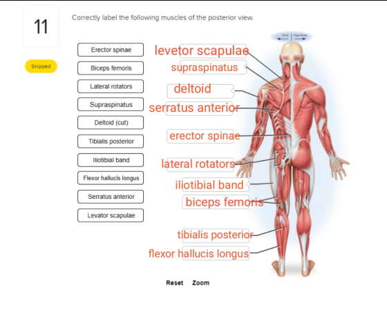
Erector spinae consists of the iliocostalis, longissimus and spinalsis muscle.
Biceps femoris is the muscle of legs. It's in the posterior aspect. In the origin it has two heads
Lateral rotators are at the hip region. They help in the rotatory movement at the hip region.
Supraspinatus is the muscle in the cuff of shoulder in the posterior aspect. It is a small muscle.
Deltoid. Is the shoulder muscle. One of the sting muscle if the 3 muscle if the shoulder. It's in the lateral aspect of the shoulder.
Tibialis posterior is in the deep posterior aspect of leg. It helps in stabilization of the leg.
Iliotibial band runs along the lateral side of the thigh. Helps in lateral knee stabilization.
Add Answer to:
Correctly label the following muscles of the posterior
view.
Correctly label the following muscles of the...
Correctly label the following muscles of the posterior view. Derec Tibialis posterior Levator scapulae lliotibial band...
 Correctly label the following muscles of the posterior view. Derec Tibialis posterior Levator scapulae lliotibial band Deltoid (cut) Biceps femoris Serratus anterior Lateral rotators Flexor hallucis longus Erector spinae Supraspinatus Reset Zoom
Correctly label the muscles of the leg. Correctly label the muscles of the leg. 57 Plantaris...
 Correctly label the muscles of the leg.
 57 Plantaris (cut) Calcaneal tendon (cut) Flexor digitorum longus Gastrocnemius (cut) Fibularis brevis Flexor hallucis longus Soleus (cut) Fibularis longus Tibialis posterior Popliteus Reset Zoom
Correctly label the muscles of the leg.
Correctly label the muscles of the leg. 57 Plantaris (cut) Calcaneal tendon (cut) Flexor digitorum longus Gastrocnemius (cut) Fibularis brevis Flexor hallucis longus Soleus (cut) Fibularis longus Tibialis posterior Popliteus Reset Zoom
Correctly label the muscles of the leg. Correctly label the muscles of the leg. Fibularis longus...
 Correctly label the muscles of the leg.
 Fibularis longus Fibularis brevis Tendon of plantaris Popliteus Flexor digitorum longus UUUUUUUUUU Plantaris Soleus Flexor hallucis longus Gastrocnemius (cut) Heads of gastrocnemius (cut) Reset Zoom
Correctly label the muscles of the leg.
Correctly label the muscles of the leg. Fibularis longus Fibularis brevis Tendon of plantaris Popliteus Flexor digitorum longus UUUUUUUUUU Plantaris Soleus Flexor hallucis longus Gastrocnemius (cut) Heads of gastrocnemius (cut) Reset Zoom
please label the matching *ctly label the muscles of the leg. Flexor digitorum longus Calcaneal tendon...
 please label the matching
*ctly label the muscles of the leg. Flexor digitorum longus Calcaneal tendon (cut) Gastrocnemius (cut) Popliteus Fibularis longus Plantaris (cut) Tibialis posterior Soleus (cut) Fibularis brevis Flexor hallucis longus
please label the matching
*ctly label the muscles of the leg. Flexor digitorum longus Calcaneal tendon (cut) Gastrocnemius (cut) Popliteus Fibularis longus Plantaris (cut) Tibialis posterior Soleus (cut) Fibularis brevis Flexor hallucis longus
please help label the diagram with the following muscles thank you. (can you please label the...
 please help label the diagram with the following muscles thank
you. (can you please label the diagram that is
given) if the muscle is not in the diagram where
would it be found
Adductor femoris Biceps femoris Caudofemoralis Extensor digitorum lateralis Flexor digitorum longus Gastrocnemius Gluteus maximus Gracilis Peroneus Rectus femoris Sartorius Semimembranosus Semitendinosus Soleus Tensor fasciae latae Tibialis anterior Vastus lateralis Vastus medialis
please help label the diagram with the following muscles thank
you. (can you please label the diagram that is
given) if the muscle is not in the diagram where
would it be found
Adductor femoris Biceps femoris Caudofemoralis Extensor digitorum lateralis Flexor digitorum longus Gastrocnemius Gluteus maximus Gracilis Peroneus Rectus femoris Sartorius Semimembranosus Semitendinosus Soleus Tensor fasciae latae Tibialis anterior Vastus lateralis Vastus medialis
the appropriate labels to their respective targets. Deltoid Trapezius Pronator teres Brachioradialis Triceps brachii Biceps brachii...
 the appropriate labels to their respective targets. Deltoid Trapezius Pronator teres Brachioradialis Triceps brachii Biceps brachii Submit Request Answer appropriate labels to their respective targets. Reset C Pectoralis minor Platysma Serratus anterior Intercostals Sternocleidomastoid Pectoralis major Sternohyoid Submit Previous Answers Request Answer rag the appropriate labels to their respective targets. Rectus femoris Vastus medialis Tibialis anterior Vastus lateralis Soleus Fibularis longus Gastrocnemius Submit Request Answer ovide Feedback eling Activity: Figure 10.5 (3 of 3) Levator scapulae Gluteus maximus Rhomboid major...
Date 1 What spinal nerve roots provide fibers to the brachial plexus? 2 Name the five...
 Date 1 What spinal nerve roots provide fibers to the brachial plexus? 2 Name the five major peripheral nerves that originate from the brachial plexus. 3 For each of the muscles listed, indicate one major action that results when this muscle contracts. Use your textbook as a resource. Biceps brachii Brachialis Deltoid Extensor carpi flexion at the elbow radialis /ulnaris) Extensor digitorum Flexor carpi radialis (radialis/ ulnaris) Flexor digitorum (superficialis/ profundus) Infraspinatus Levator scapulae Rhomboideus Triceps brachii Serratus anterior Supraspinatus
238 EXERCISE TEN - Muscular System 16. Which is not a flexor of the forearm? (a)...
 238 EXERCISE TEN - Muscular System 16. Which is not a flexor of the forearm? (a) biceps brachii (b) brachialis (c) brachioradialis (d) triceps brachii 17. The abductor pollicis brevis, opponeus pollicis, flexor pollicis brevis, and adductor polli- cis are all components of the (a) thenar eminence (b) midpalmar muscles (C) hypothenar eminence (d) erector spinae 18. Which muscle is not involved in moving the vertebral column? (a) splenius (b) longis- simus (c) spinalis (d) sartorius 19. Which muscle flexes...
Correctly label the following muscles of the anterior view. Superficial | Deep Frontalis Brachiordialis Vastus lateralis...
 Correctly label the following muscles of the anterior
view.
Superficial | Deep Frontalis Brachiordialis Vastus lateralis Biceps brachii External abdominal oblique Fibularis longus Platysma Adductor longus Tensor fasciae latae Pectoralis major
Correctly label the following muscles of the anterior
view.
Superficial | Deep Frontalis Brachiordialis Vastus lateralis Biceps brachii External abdominal oblique Fibularis longus Platysma Adductor longus Tensor fasciae latae Pectoralis major
fill in the blank boxes all 6 Correctly label the anterior muscles of the thigh. 35...
 fill in the blank boxes all 6
Correctly label the anterior muscles of the thigh. 35 Quadriceps femoris: Vastus medialis Patellar ligament Quadriceps femoris: Vastus lateralis Quadriceps femoris: Vastus intermedius Quadriceps femoris tendon Patella Reset Zoom cles of the thigh. 35
fill in the blank boxes all 6
Correctly label the anterior muscles of the thigh. 35 Quadriceps femoris: Vastus medialis Patellar ligament Quadriceps femoris: Vastus lateralis Quadriceps femoris: Vastus intermedius Quadriceps femoris tendon Patella Reset Zoom cles of the thigh. 35
 Correctly label the following muscles of the posterior view. Derec Tibialis posterior Levator scapulae lliotibial band Deltoid (cut) Biceps femoris Serratus anterior Lateral rotators Flexor hallucis longus Erector spinae Supraspinatus Reset Zoom
 Correctly label the muscles of the leg.
 57 Plantaris (cut) Calcaneal tendon (cut) Flexor digitorum longus Gastrocnemius (cut) Fibularis brevis Flexor hallucis longus Soleus (cut) Fibularis longus Tibialis posterior Popliteus Reset Zoom
Correctly label the muscles of the leg.
Correctly label the muscles of the leg. 57 Plantaris (cut) Calcaneal tendon (cut) Flexor digitorum longus Gastrocnemius (cut) Fibularis brevis Flexor hallucis longus Soleus (cut) Fibularis longus Tibialis posterior Popliteus Reset Zoom
 Correctly label the muscles of the leg.
 Fibularis longus Fibularis brevis Tendon of plantaris Popliteus Flexor digitorum longus UUUUUUUUUU Plantaris Soleus Flexor hallucis longus Gastrocnemius (cut) Heads of gastrocnemius (cut) Reset Zoom
Correctly label the muscles of the leg.
Correctly label the muscles of the leg. Fibularis longus Fibularis brevis Tendon of plantaris Popliteus Flexor digitorum longus UUUUUUUUUU Plantaris Soleus Flexor hallucis longus Gastrocnemius (cut) Heads of gastrocnemius (cut) Reset Zoom
 please label the matching
*ctly label the muscles of the leg. Flexor digitorum longus Calcaneal tendon (cut) Gastrocnemius (cut) Popliteus Fibularis longus Plantaris (cut) Tibialis posterior Soleus (cut) Fibularis brevis Flexor hallucis longus
please label the matching
*ctly label the muscles of the leg. Flexor digitorum longus Calcaneal tendon (cut) Gastrocnemius (cut) Popliteus Fibularis longus Plantaris (cut) Tibialis posterior Soleus (cut) Fibularis brevis Flexor hallucis longus
 please help label the diagram with the following muscles thank
you. (can you please label the diagram that is
given) if the muscle is not in the diagram where
would it be found
Adductor femoris Biceps femoris Caudofemoralis Extensor digitorum lateralis Flexor digitorum longus Gastrocnemius Gluteus maximus Gracilis Peroneus Rectus femoris Sartorius Semimembranosus Semitendinosus Soleus Tensor fasciae latae Tibialis anterior Vastus lateralis Vastus medialis
please help label the diagram with the following muscles thank
you. (can you please label the diagram that is
given) if the muscle is not in the diagram where
would it be found
Adductor femoris Biceps femoris Caudofemoralis Extensor digitorum lateralis Flexor digitorum longus Gastrocnemius Gluteus maximus Gracilis Peroneus Rectus femoris Sartorius Semimembranosus Semitendinosus Soleus Tensor fasciae latae Tibialis anterior Vastus lateralis Vastus medialis
 the appropriate labels to their respective targets. Deltoid Trapezius Pronator teres Brachioradialis Triceps brachii Biceps brachii Submit Request Answer appropriate labels to their respective targets. Reset C Pectoralis minor Platysma Serratus anterior Intercostals Sternocleidomastoid Pectoralis major Sternohyoid Submit Previous Answers Request Answer rag the appropriate labels to their respective targets. Rectus femoris Vastus medialis Tibialis anterior Vastus lateralis Soleus Fibularis longus Gastrocnemius Submit Request Answer ovide Feedback eling Activity: Figure 10.5 (3 of 3) Levator scapulae Gluteus maximus Rhomboid major...
 Date 1 What spinal nerve roots provide fibers to the brachial plexus? 2 Name the five major peripheral nerves that originate from the brachial plexus. 3 For each of the muscles listed, indicate one major action that results when this muscle contracts. Use your textbook as a resource. Biceps brachii Brachialis Deltoid Extensor carpi flexion at the elbow radialis /ulnaris) Extensor digitorum Flexor carpi radialis (radialis/ ulnaris) Flexor digitorum (superficialis/ profundus) Infraspinatus Levator scapulae Rhomboideus Triceps brachii Serratus anterior Supraspinatus
 238 EXERCISE TEN - Muscular System 16. Which is not a flexor of the forearm? (a) biceps brachii (b) brachialis (c) brachioradialis (d) triceps brachii 17. The abductor pollicis brevis, opponeus pollicis, flexor pollicis brevis, and adductor polli- cis are all components of the (a) thenar eminence (b) midpalmar muscles (C) hypothenar eminence (d) erector spinae 18. Which muscle is not involved in moving the vertebral column? (a) splenius (b) longis- simus (c) spinalis (d) sartorius 19. Which muscle flexes...
 Correctly label the following muscles of the anterior
view.
Superficial | Deep Frontalis Brachiordialis Vastus lateralis Biceps brachii External abdominal oblique Fibularis longus Platysma Adductor longus Tensor fasciae latae Pectoralis major
Correctly label the following muscles of the anterior
view.
Superficial | Deep Frontalis Brachiordialis Vastus lateralis Biceps brachii External abdominal oblique Fibularis longus Platysma Adductor longus Tensor fasciae latae Pectoralis major
 fill in the blank boxes all 6
Correctly label the anterior muscles of the thigh. 35 Quadriceps femoris: Vastus medialis Patellar ligament Quadriceps femoris: Vastus lateralis Quadriceps femoris: Vastus intermedius Quadriceps femoris tendon Patella Reset Zoom cles of the thigh. 35
fill in the blank boxes all 6
Correctly label the anterior muscles of the thigh. 35 Quadriceps femoris: Vastus medialis Patellar ligament Quadriceps femoris: Vastus lateralis Quadriceps femoris: Vastus intermedius Quadriceps femoris tendon Patella Reset Zoom cles of the thigh. 35
Most questions answered within 3 hours.
-
Where is the error in this code sequence?
String s1 = "Hello";
String s2 = "ello";...
asked 11 months ago -
Financial data for Joel de Paris, Inc., for last year
follow:
Joel de Paris, Inc.
Balance...
asked 11 months ago -
Consider this reaction:
Al2(SO4)3 (aq)+ BaCl3
(aq) Al2Cl6 (aq)- +
3BaSO4(s) . What is the...
asked 11 months ago -
Suppose that Savneet is considering increasing her
recent random sample from 20 car rentals to 40...
asked 11 months ago -
Trucks arrive at an unloading terminal at an average rate of 120
per hour.
Trucks arrive...
asked 11 months ago -
Why are methanol and ethanol completely soluble in water while
octanol is not very little soluble....
asked 11 months ago -
A facilities manager at a university reads in a research report
that the mean amount of...
asked 11 months ago -
When the CuSO4 is rehydrated by adding water to the anhydrous
compound, is this an endothermic...
asked 11 months ago -
A ray of sunlight is passing from diamond into crown glass; the
angle of incidence is...
asked 11 months ago -
A block of mass 0.249 kg is placed on top of a light, vertical
spring of...
asked 11 months ago -
how do the kidneys compensate in the presences of acidosis
a) trigger hyperventilate
b) reserve acid...
asked 11 months ago -
Question 501 pts
The rental rate of capital to the firm increases. Which of the
following...
asked 11 months ago