
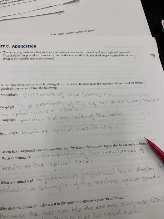
dorsal root I vou expect to find myelinated axons? rt C: Application Women giving birth are often given an anesthetic medication into the epidural space (epidural anesthesia), Occasionally this procedure causes a tear in the dura mater. What do you think might happen if this occurs? What is the possible risk to the woman? Sometimes the spinal cord can he damaged in an accident. Depending on the location and severity of the iur, and louay body) сапусо on spinalinodiscate. inot pinal Cord damas uadriplegia: patient has a suspected case of meningitis. The physician orders a spinal tap so that he can make a diagno on o the wrem brones What is meningitis? i brain or the neinal cofa What is a spinal tap ayre cature pregorYmeơ on a dockor a cerebral spinali Why does the physician order a test in the spine to diagnosis a problem in the head? causs the lest can hdp dhe de hora dlianox dissr inpr the spinal cord
Homework Answers
Epidural anesthesia is a method of administration of anesthesia into epidural space . Epidural space is located between ligamentum flavum and dura matter. The space is about 3 -5 mm and is easy to get accidental puncture of dura matter. The anesthetia is administered into lumbar epidural space from there it diffuse into dura and into subarachinoid space. The accidental puncture may cause CSF to leak out into epidural space and reduction in pressure of CSF which cause post dural puncture headache (PDPH). This is severe and last from days to weeks and is characterised by postdural exacerbation when patients head is raised above normal lying position . Epidural blood patch is a treatment method where patient's own blood is given to the dural space where it clots and seals the leak.
Add Answer to:
Part B: Working With Histology Slides Trachea Slide Now that you have some experience with the hi...
PROCEDURE Exercise 20-2 Lymphatic Organ Histology Microscopy 1 Obtain prepared slides of the sple...
 PROCEDURE Exercise 20-2 Lymphatic Organ Histology Microscopy 1 Obtain prepared slides of the spleen, a lymph node, and the small intestine (be certain that your slide is labeled the ilee) MATERIALS u Spleen section slide Light microscope J Lymph node slide 2 Use your colored pencils to draw what you see in the fiel Colored pencils of view and label your drawing with the terms below. Peyer's patch slide Spleen 1. Red pulp 2. White pulp In this exercise, you...
 PROCEDURE Exercise 20-2 Lymphatic Organ Histology Microscopy 1 Obtain prepared slides of the spleen, a lymph node, and the small intestine (be certain that your slide is labeled the ilee) MATERIALS u Spleen section slide Light microscope J Lymph node slide 2 Use your colored pencils to draw what you see in the fiel Colored pencils of view and label your drawing with the terms below. Peyer's patch slide Spleen 1. Red pulp 2. White pulp In this exercise, you...
Most questions answered within 3 hours.
-
Where is the error in this code sequence?
String s1 = "Hello";
String s2 = "ello";...
asked 11 months ago -
Financial data for Joel de Paris, Inc., for last year
follow:
Joel de Paris, Inc.
Balance...
asked 11 months ago -
Consider this reaction:
Al2(SO4)3 (aq)+ BaCl3
(aq) Al2Cl6 (aq)- +
3BaSO4(s) . What is the...
asked 11 months ago -
Suppose that Savneet is considering increasing her
recent random sample from 20 car rentals to 40...
asked 11 months ago -
Trucks arrive at an unloading terminal at an average rate of 120
per hour.
Trucks arrive...
asked 11 months ago -
Why are methanol and ethanol completely soluble in water while
octanol is not very little soluble....
asked 11 months ago -
A facilities manager at a university reads in a research report
that the mean amount of...
asked 11 months ago -
When the CuSO4 is rehydrated by adding water to the anhydrous
compound, is this an endothermic...
asked 11 months ago -
A ray of sunlight is passing from diamond into crown glass; the
angle of incidence is...
asked 11 months ago -
A block of mass 0.249 kg is placed on top of a light, vertical
spring of...
asked 11 months ago -
how do the kidneys compensate in the presences of acidosis
a) trigger hyperventilate
b) reserve acid...
asked 11 months ago -
Question 501 pts
The rental rate of capital to the firm increases. Which of the
following...
asked 11 months ago