
Homework Answers
Add Answer to:
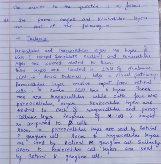
QUESTION 86 The parvo-magno- and koniocellular layers are part of the thalamus the retina the smatosensory...
Question 47 Describe a visual pathway from the retina to the visual cortex of the occipital...
 Question 47 Describe a visual pathway from the retina to the visual cortex of the occipital lobe. Include the terms in the textbox photoreceptors Irods optic tract Icones retina thalamus ganglion cells optic disc visual cortex of occipital lobe optic nerve optic chiasm 1.! HTML Editor BIA-N-I E *** 12pt - 2 ce to search
Jestion Completion Status: QUESTION 1 Information from the left visual field travels through the eye, to...
 Jestion Completion Status: QUESTION 1 Information from the left visual field travels through the eye, to the left nasal retina, via the left optic nerve through the optic chiasma, to the right lateral geniculate nucleus of the to the primary visual cortex of the right occipital lobe. QUESTION 2 The process in which visual information from the primary visual cortex combines with other information in additional repons of the brain refers to what step of nervous system processing? O a....
Light first passes through the _____________, a transparent covering of the eye, then through the _____________,...
Light first passes through the _____________, a transparent covering of the eye, then through the _____________, the opening in the iris. Then the light passes through the _____________, which changes shape to focus the light so that it properly hits the retina. The light travels through the vitreous humor of the eye and then strikes the _____________, layers of cells in the back of the eye. The deepest layer of the retina is made of ____________, of which there are...
The Pathways of Light and Visual Information Select from the following terms to fill in the...
The Pathways of Light and Visual Information Select from the following terms to fill in the blanks in the passage below. Use each term once. bipolar cones cornea ganglion optic nerve lens rods retina transduce optic chiasm occipital pupil pattern photoreceptors Light first passes through the _____________, a transparent covering of the eye, then through the _____________, the opening in the iris. Then the light passes through the _____________, which changes shape to focus the light so that it properly...
these two, pls. Visually guided movement is possible in the absence of conscious visual perception, a...
 these two, pls.
Visually guided movement is possible in the absence of conscious visual perception, a phenomenon referred to as "blindsight". Similarly, fearful images can elicit emotional responses in the absence of their conscious perception. True or false the circuit that allows for emotional responses, to potentially dangerous visual stimuli, in the absence of the conscious perception of that visual stimull is as follows: retina->lateral geniculate nucleus (thalamus)->primary visual cortex->amygdala->autonomic nervous system O True O False You are an expert...
these two, pls.
Visually guided movement is possible in the absence of conscious visual perception, a phenomenon referred to as "blindsight". Similarly, fearful images can elicit emotional responses in the absence of their conscious perception. True or false the circuit that allows for emotional responses, to potentially dangerous visual stimuli, in the absence of the conscious perception of that visual stimull is as follows: retina->lateral geniculate nucleus (thalamus)->primary visual cortex->amygdala->autonomic nervous system O True O False You are an expert...
4. The primary relay nucleus between the retina and the cerebral cortex is called the A)...
 4. The primary relay nucleus between the retina and the cerebral cortex is called the A) superior colliculus C) suprachiasmatic nucleus. B) lateral geniculate nucleus. D) medial geniculate nucleus. Part 2. The link below contains important information about structure and function in visual processing Select the link. http://www.sumanasinc.com/webcontent/animations/content/receptivefields.html. View the content. For each of the following quiz questions, highlight the letter corresponding to the correct answer 5. The optic nerves, which are made up the axons of the the brain....
QUESTION 25 The right and left cerebral hemispheres are connected by A The thalamus B.the best...
 QUESTION 25 The right and left cerebral hemispheres are connected by A The thalamus B.the best The basalgia D. The corpus callosum QUESTION 26 Hydrocephalus in canned by A a blockage in the normal flow of CSF (cerebrospinal fluid) B. Inflammation of the time rounding the brain C. cutting the corpus callosum Desis is the prefrontal cortex QUESTION 27 In which part of the brain is the thalamus located? A the forebrain B. the midbrain C the hindbrain D. none...
l alEW Backtracking. Changes to the answer after submission are prohibited. Remaining Time:6 minutes, 08 seconds....
 l alEW Backtracking. Changes to the answer after submission are prohibited. Remaining Time:6 minutes, 08 seconds. Question Completion Status: ) Moving to the next question prevents changes to this answer Question 8 Sarting with activation of receptors, what is the correct path visual information takes to travel from the retina to the primary visual cortex? Rods; bipolar cells: retinal ganglion cells; optic nerve; optic tract, lateral geniculate nucleus Cones; retinal ganglion cells; optic tract; optic nerve; thalamus; hypothalamus Retinal ganglion...
60. Double-opponent cells are first found in the a, retina. b. optic nerve. c. optic chiasm....
 60. Double-opponent cells are first found in the a, retina. b. optic nerve. c. optic chiasm. d. LGN. e. primary visual cortex 61. The diagram below illustrates the depth cue. a. motion parallax b. aerial perspective c. linear perspective d. accommodation e. convergence 62. A(n) is a visual image seen after the stimulus has been removed. a. adapting stimulus b. afterimage c. neutral point d. metamer e, hallucination 63. What is the optic array? 64. Parallel lines in the world...
This is the question:::: discuss what visual information is processed by the retina and by simple...
 This is the question:::: discuss what visual information is
processed by the retina and by simple and complex cells in V1, and
what the differences are between these levels of information
processing.
Question 17 3 pts Use several paragraphs to discuss what visual information is processed by the retina and by simple and complex cells in V1, and what the differences are between these levels of information processing. HTML Editor B 3 3 := = IVA - A - IX...
This is the question:::: discuss what visual information is
processed by the retina and by simple and complex cells in V1, and
what the differences are between these levels of information
processing.
Question 17 3 pts Use several paragraphs to discuss what visual information is processed by the retina and by simple and complex cells in V1, and what the differences are between these levels of information processing. HTML Editor B 3 3 := = IVA - A - IX...
 Question 47 Describe a visual pathway from the retina to the visual cortex of the occipital lobe. Include the terms in the textbox photoreceptors Irods optic tract Icones retina thalamus ganglion cells optic disc visual cortex of occipital lobe optic nerve optic chiasm 1.! HTML Editor BIA-N-I E *** 12pt - 2 ce to search
 Jestion Completion Status: QUESTION 1 Information from the left visual field travels through the eye, to the left nasal retina, via the left optic nerve through the optic chiasma, to the right lateral geniculate nucleus of the to the primary visual cortex of the right occipital lobe. QUESTION 2 The process in which visual information from the primary visual cortex combines with other information in additional repons of the brain refers to what step of nervous system processing? O a....
 these two, pls.
Visually guided movement is possible in the absence of conscious visual perception, a phenomenon referred to as "blindsight". Similarly, fearful images can elicit emotional responses in the absence of their conscious perception. True or false the circuit that allows for emotional responses, to potentially dangerous visual stimuli, in the absence of the conscious perception of that visual stimull is as follows: retina->lateral geniculate nucleus (thalamus)->primary visual cortex->amygdala->autonomic nervous system O True O False You are an expert...
these two, pls.
Visually guided movement is possible in the absence of conscious visual perception, a phenomenon referred to as "blindsight". Similarly, fearful images can elicit emotional responses in the absence of their conscious perception. True or false the circuit that allows for emotional responses, to potentially dangerous visual stimuli, in the absence of the conscious perception of that visual stimull is as follows: retina->lateral geniculate nucleus (thalamus)->primary visual cortex->amygdala->autonomic nervous system O True O False You are an expert...
 4. The primary relay nucleus between the retina and the cerebral cortex is called the A) superior colliculus C) suprachiasmatic nucleus. B) lateral geniculate nucleus. D) medial geniculate nucleus. Part 2. The link below contains important information about structure and function in visual processing Select the link. http://www.sumanasinc.com/webcontent/animations/content/receptivefields.html. View the content. For each of the following quiz questions, highlight the letter corresponding to the correct answer 5. The optic nerves, which are made up the axons of the the brain....
 QUESTION 25 The right and left cerebral hemispheres are connected by A The thalamus B.the best The basalgia D. The corpus callosum QUESTION 26 Hydrocephalus in canned by A a blockage in the normal flow of CSF (cerebrospinal fluid) B. Inflammation of the time rounding the brain C. cutting the corpus callosum Desis is the prefrontal cortex QUESTION 27 In which part of the brain is the thalamus located? A the forebrain B. the midbrain C the hindbrain D. none...
 l alEW Backtracking. Changes to the answer after submission are prohibited. Remaining Time:6 minutes, 08 seconds. Question Completion Status: ) Moving to the next question prevents changes to this answer Question 8 Sarting with activation of receptors, what is the correct path visual information takes to travel from the retina to the primary visual cortex? Rods; bipolar cells: retinal ganglion cells; optic nerve; optic tract, lateral geniculate nucleus Cones; retinal ganglion cells; optic tract; optic nerve; thalamus; hypothalamus Retinal ganglion...
 60. Double-opponent cells are first found in the a, retina. b. optic nerve. c. optic chiasm. d. LGN. e. primary visual cortex 61. The diagram below illustrates the depth cue. a. motion parallax b. aerial perspective c. linear perspective d. accommodation e. convergence 62. A(n) is a visual image seen after the stimulus has been removed. a. adapting stimulus b. afterimage c. neutral point d. metamer e, hallucination 63. What is the optic array? 64. Parallel lines in the world...
 This is the question:::: discuss what visual information is
processed by the retina and by simple and complex cells in V1, and
what the differences are between these levels of information
processing.
Question 17 3 pts Use several paragraphs to discuss what visual information is processed by the retina and by simple and complex cells in V1, and what the differences are between these levels of information processing. HTML Editor B 3 3 := = IVA - A - IX...
This is the question:::: discuss what visual information is
processed by the retina and by simple and complex cells in V1, and
what the differences are between these levels of information
processing.
Question 17 3 pts Use several paragraphs to discuss what visual information is processed by the retina and by simple and complex cells in V1, and what the differences are between these levels of information processing. HTML Editor B 3 3 := = IVA - A - IX...
Most questions answered within 3 hours.
-
Where is the error in this code sequence?
String s1 = "Hello";
String s2 = "ello";...
asked 10 months ago -
Financial data for Joel de Paris, Inc., for last year
follow:
Joel de Paris, Inc.
Balance...
asked 10 months ago -
Consider this reaction:
Al2(SO4)3 (aq)+ BaCl3
(aq) Al2Cl6 (aq)- +
3BaSO4(s) . What is the...
asked 10 months ago -
Suppose that Savneet is considering increasing her
recent random sample from 20 car rentals to 40...
asked 10 months ago -
Trucks arrive at an unloading terminal at an average rate of 120
per hour.
Trucks arrive...
asked 10 months ago -
Why are methanol and ethanol completely soluble in water while
octanol is not very little soluble....
asked 10 months ago -
A facilities manager at a university reads in a research report
that the mean amount of...
asked 10 months ago -
When the CuSO4 is rehydrated by adding water to the anhydrous
compound, is this an endothermic...
asked 10 months ago -
A ray of sunlight is passing from diamond into crown glass; the
angle of incidence is...
asked 10 months ago -
A block of mass 0.249 kg is placed on top of a light, vertical
spring of...
asked 10 months ago -
how do the kidneys compensate in the presences of acidosis
a) trigger hyperventilate
b) reserve acid...
asked 10 months ago -
Question 501 pts
The rental rate of capital to the firm increases. Which of the
following...
asked 10 months ago