What is happening in the eye? 1. Draw the sensory receptors in the eye 2. Draw...
What is happening in the eye?
1. Draw the sensory receptors in the eye
2. Draw what an action potential looks like in a neuron (intercellular communication) Note: go through all the steps to show how membrane potential changes from the beginning to the end of an action potential
Homework Answers

Look at this picture from standard textbook.
Now, The rod and the cone cells are the sensory receptors in the eye.
Rods are responsible for black and white vision and are more active at night during low levels of light. Your cones are for color vision and are used during the day when light is ample.
Let's look closely at these receptors.

Cone cells are somewhat shorter than rods, but wider and tapered, and are much less numerous than rods in most parts of the retina, but greatly outnumber rods in the fovea. Structurally, cone cells have a cone-like shape at one end where a pigment filters incoming light, giving them their different response curves. They are typically 40–50 µm long, and their diameter varies from 0.5 to 4.0 µm, being smallest and most tightly packed at the center of the eye at the fovea. The S cone spacing is slightly larger than the others.
Like rods, each cone cell has a synaptic terminal, an inner segment, and an outer segment as well as an interior nucleus and various mitochondria. The synaptic terminal forms a synapse with a neuron such as a bipolar cell. The inner and outer segments are connected by a cilium. The inner segment contains organelles and the cell's nucleus, while the outer segment, which is pointed toward the back of the eye, contains the light-absorbing materials.
Unlike rods, the outer segments of cones have invaginations of their cell membranes that create stacks of membranous disks. Photopigments exist as transmembrane proteins within these disks, which provide more surface area for light to affect the pigments. In cones, these disks are attached to the outer membrane, whereas they are pinched off and exist separately in rods. Neither rods nor cones divide, but their membranous disks wear out and are worn off at the end of the outer segment, to be consumed and recycled. by phagocytic cells.
Rods are a little longer and leaner than cones but have the same basic structure. Opsin-containing disks lie at the end of the cell adjacent to the retinal pigment epithelium, which in turn is attached to the inside of the eye. The stacked-disc structure of the detector portion of the cell allows for very high efficiency. Rods are much more common than cones, with about 120 million rod cells compared to 6 to 7 million cone cells.
Like cones, rod cells have a synaptic terminal, an inner segment, and an outer segment. The synaptic terminal forms a synapse with another neuron, usually a bipolar cell or a horizontal cell. The inner and outer segments are connected by a cilium, which lines the distal segment.The inner segment contains organelles and the cell's nucleus, while the rod outer segment (abbreviated to ROS), which is pointed toward the back of the eye, contains the light-absorbing materials.
A human rod cell is about 2 microns in diameter and 100 microns long. Rods are not all morphologically the same; in mice, rods close to the outer plexiform synaptic layer display a reduced length due to a shortened synaptic terminal.
How action potential looks like?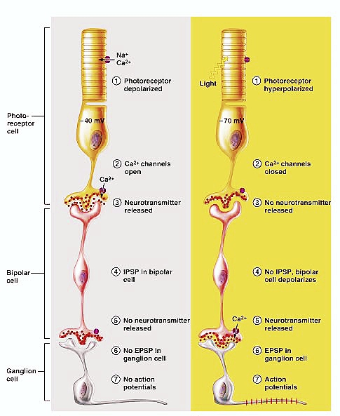
Without Light. With Light.
It clearly depicts the entire proces till the generation of action potential.
In the absence of light, the photoreceptors are depolarized to a membrane resting potential of -40mV. Light will hyperpolarize the plasma membrane of the photoreceptor to -70mV. This stimulus-induced hyperpolarization is a distinctive characteristic of the photoreceptor response, as many other neuronal types depolarize when stimulated.
A key second messenger molecule responsible for maintaining a depolarized rest state in photoreceptors is the nucleotide cyclic guanosine 3’-5’ monophospate (cGMP). High cGMP levels keep cGMP-gated ion channels in the open state and allow them to pass an inward Na+ current.
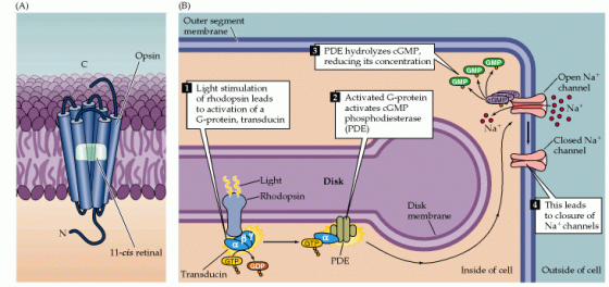
Phototransduction involves three main biochemical events:
Light entering the eye activates the opsin molecules in the photoreceptors
Upon photon absorption, 11-cis-retinal undergoes an isomerization to the all-trans form, causing a conformational change in the rhodopsin. The activated rhodopsin is called metarhodopsin II.
The precursor for 11-cis-retinal is all-trans-retinol (vitamin A). A diet rich in vitamin A is crucial for vision, since vitamin A cannot be synthesized by humans.
Activated rhodopsin causes a reduction in the cGMP intracellular concentration.
The cytoplasmic cGMP levels are controlled by cGMP phosphodiesterase, an enzyme that breaks down cGMP. In the dark, the activity of this enzyme is relatively weak. When the photoreceptor is exposed to light, metarhodopsin II stimulates the activity of cGMP phosphodiesterase via transducin, a G protein. GDP-bound inactive transducin will exchange GDP for GTP following interaction with activated rhodopsin. GTP-bound active transducin will increase the activity of cGMP phosphodiesterase. The result is decreased levels of cGMP in the cytoplasm.
The photoreceptor is hyperpolarized following exposure to light
Decreased levels of cGMP cause the closing of cGMP-gated ion channels which will lead to membrane hyperpolarization.
Termination of the phototransduction cascade
The light response is terminated by several mechanisms.
(1) Inactivation of rhodopsin occurs through phosphorylation by the opsin kinase, followed by the binding of arrestin to phosphorylated rhodopsin.
(2) Inactivation of transducin occurs through the hydrolysis of bound GTP to GDP (Tα-GTP to Tα-GDP) via an intrinsic GTPase activity that is accelerated by the GTPase activating protein RGS9 (regulator of G-protein signaling).
(3) Inactivation of phosphodiesterase (PDE) is coupled to the inactivation of transducin. Inactivated transducin (Tα-GDP) dissociates from PDE, resulting in a cessation of PDE-mediated cGMP hydrolysis.
(4) Activation of guanylate cyclase by guanylate cyclase activating protein (GCAP) restores cGMP levels and thus promotes the re-opening of cGMP-gated channels.
Amplification in the phototransduction cascade
The activation of a single rhodopsin by a single photon is sufficient to cause a significant change in the membrane conductance. This is possible due to amplification steps present in the transduction cascade.
A single photoactivated rhodopsin catalyses the activation of 500 transducin molecules. Each transducing can stimulate one cGMP phosphodiesterase molecule and each cGMP phosphodiesterase molecule can break down 10^3 molecules of cGMP per second. Therefore, a single activated rhodopsin can cause the hydrolysis of more than 10^5 molecules of cGMP per second.
From here on,

Extracranial
The optic nerve is formed by the convergence of axons from the retinal ganglion cells. These cells in turn receive impulses from the photoreceptors of the eye (the rods and cones).
After its formation, the nerve leaves the bony orbit via the optic canal, a passageway through the sphenoid bone. It enters the cranial cavity, running along the surface of the middle cranial fossa (in close proximity to the pituitary gland).
Intracranial (The Visual Pathway)
Within the middle cranial fossa, the optic nerves from each eye unite to form the optic chiasm. At the chiasm, fibres from the nasal (medial) half of each retina cross over to the contralateral optic tract, while fibres from the temporal (lateral) halves remain ipsilateral:
- Left optic tract – contains fibres from the left temporal (lateral) retina, and the right nasal (medial) retina.
- Right optic tract – contains fibres from the right temporal retina, and the left nasal retina.
Each optic tract travels to its corresponding cerebral hemisphere to reach the lateral geniculate nucleus (LGN), a relay system located in the thalamus; the fibres synapse here.
Axons from the LGN then carry visual information via a pathway known as the optic radiation. The pathway itself can be divided into:
- Upper optic radiation – carries fibres from the superior retinal quadrants (corresponding to the inferior visual field quadrants). It travels through the parietal lobe to reach the visual cortex.
- Lower optic radiation – carries fibres from the inferior retinal quadrants (corresponding to the superior visual field quadrants). It travels through the temporal lobe, via a pathway known as Meyers’ loop, to reach the visual cortex.
Once at the visual cortex, the brain processes the sensory data and responds appropriately.
Add Answer to:
What is happening in the eye?
1. Draw the sensory receptors in the eye
2. Draw...
1. The following figure shows that a sensory neuron detects the stimulation and sends a signal...
 1. The following figure shows that a sensory neuron detects the stimulation and sends a signal to a motor neuron. How does the sensory neuron pass a signal to the motor neuron? Start your answer from action potential propagation along the sensory axon. You want to include detailed steps of neurotransmitter release in your answer. (12 points) 2. What is the neurotransmitter used by the sensory neuron? (1 point) Sensory afferent Muscle sensory receptor Extensor muscle Motorefferent
2. Curare blocks acetylcholine receptors at the motor end plate. This would result in: (1) Inability...
 2. Curare blocks acetylcholine receptors at the motor end plate. This would result in: (1) Inability of the muscle fiber to respond to nervous stimulation (2) Increased muscle stimulation (3) Increased actylcholinesterase production (4) Lack of calcium uptake by the muscle fiber 3. The structure that is analogous to the Zline of skeletal muscle is the __ of smooth muscle. The structure that is analogous to troponin of skeletal muscle is _ muscle is ___ of smooth muscle. 4. Draw...
4. Draw a synapse between 2 neurons. Label the following: Presynaptic neuron, Postsynaptic neuron, Synaptic vesicles,...
 4. Draw a synapse between 2 neurons. Label the following: Presynaptic neuron, Postsynaptic neuron, Synaptic vesicles, Voltage-regulated calcium channel, Chemical-regulated sodium channel. 5. Draw a diagram of the preganglionic neuron, postganglionic neuron, and effector for both Sympathetic Nervous System and Parasympathetic Nervous System. indicate which neurotransmitter is released by each neuron and label the receptors at all locations for the neurotransmitter. 6. Which cells have a resting membrane potential? Which cells can have an action potential? 7. Circle which of...
For each multiple choice, what is the answer and why? 5. Myelin A) is only found...
 For
each multiple choice, what is the answer and why?
5. Myelin A) is only found in the peripheral nervous system B) is secreted from the axon terminals of autonomic neurons. C) is a carbohydrate within the membranes of some neuroglia. D) is a chemical present in the plasma membrane of neurons. E) influences the rate of conduction of an electrical signal down an axon. 6. Which of the following organs/structures is (are) innervated by somatic motor nerves? (only ONE...
For
each multiple choice, what is the answer and why?
5. Myelin A) is only found in the peripheral nervous system B) is secreted from the axon terminals of autonomic neurons. C) is a carbohydrate within the membranes of some neuroglia. D) is a chemical present in the plasma membrane of neurons. E) influences the rate of conduction of an electrical signal down an axon. 6. Which of the following organs/structures is (are) innervated by somatic motor nerves? (only ONE...
Please answer as many as possible NERVOUS TISSUE Draw a diagram that shows the components and...
 Please answer as many as possible
NERVOUS TISSUE Draw a diagram that shows the components and interrelationships between the divisions of the nervous system. Sketch a multipolar neuron and label its major parts Explain the differences between unipolar, bipolar, and multipolar neurons. Explain the differences between sensory, association (interneuron), and motor neurons. How is a glial cell different from neuron? What is myelin? How is it formed differently in the PNS compared to the CNS? Explain how an excitable cell...
Please answer as many as possible
NERVOUS TISSUE Draw a diagram that shows the components and interrelationships between the divisions of the nervous system. Sketch a multipolar neuron and label its major parts Explain the differences between unipolar, bipolar, and multipolar neurons. Explain the differences between sensory, association (interneuron), and motor neurons. How is a glial cell different from neuron? What is myelin? How is it formed differently in the PNS compared to the CNS? Explain how an excitable cell...
Membrane-bound receptors are an important component facilitating some types of cellular communication; however, not all signal-receptor...
Membrane-bound receptors are an important component facilitating some types of cellular communication; however, not all signal-receptor binding occurs outside the cell membrane. Some signal molecules are able to pass through the plasma membrane, and their receptors are intracellular. In a theoretical lab study, scientists work to design a lab experiment that would inhibit some molecules’ ability to bind with their intracellular receptors. (a) Draw conclusions about the structure and function of membrane proteins. (b) Identify some molecules that are able...
You are recording from a neuron that has without current injection a tonic firing pattern of...
 You are recording from a neuron that has without current
injection a tonic firing pattern of action potentials. While you
inject a long pulse of a depolarizing current, the spikes that the
cell produces are first quite large, but become wider over time,
their amplitude decreases, and eventually there is no response any
more. It looks like of like this:
For a moment you then hyperpolarize the cell. Then you
depolarize the cell again, and you see the same thing...
You are recording from a neuron that has without current
injection a tonic firing pattern of action potentials. While you
inject a long pulse of a depolarizing current, the spikes that the
cell produces are first quite large, but become wider over time,
their amplitude decreases, and eventually there is no response any
more. It looks like of like this:
For a moment you then hyperpolarize the cell. Then you
depolarize the cell again, and you see the same thing...
What is happening in the ear? 1. Draw the structures of the ear (the outer, middle,...
What is happening in the ear? 1. Draw the structures of the ear (the outer, middle, and inner ear) 2. Draw how signal transduction happens in the cochlea 3. Draw how information is organized along the basilar membrane
KEY TERMS March the numbered form with the definition that is it be definition the responding...
 KEY TERMS March the numbered form with the definition that is it be definition the responding sumber in front of the appropriate 40. excitatory postsynaptic potential 21. resting potential 22 on leakage channels 23. equilibrium potential -24 graded potential 25. gated ion channels 26. depolarization 27. hyperpolarization Sensory neurons 2. central nervous system 3. motor neurons 4. intercurons 5. peripheral nervous system 6. somatic motor neurons *7. autonomic motor neurons 8. sympathetic 9. parasympathetic 10. cell body all dendrites 12....
please answer - What is the purpose of an EEG? What sends the signal! Wildt Which...
 please answer
- What is the purpose of an EEG? What sends the signal! Wildt Which parts of the brain are involved in movement? Practice Questions uestions. These questions are for practice. All possible content may not be represented in this subset of question Dita 1. Jn which area of the cerebrum is the visual cortex located? 2. The is thought to be the involved in learning and memory. 3. The specialization of each cerebral hemisphere for certain functions is...
please answer
- What is the purpose of an EEG? What sends the signal! Wildt Which parts of the brain are involved in movement? Practice Questions uestions. These questions are for practice. All possible content may not be represented in this subset of question Dita 1. Jn which area of the cerebrum is the visual cortex located? 2. The is thought to be the involved in learning and memory. 3. The specialization of each cerebral hemisphere for certain functions is...
 1. The following figure shows that a sensory neuron detects the stimulation and sends a signal to a motor neuron. How does the sensory neuron pass a signal to the motor neuron? Start your answer from action potential propagation along the sensory axon. You want to include detailed steps of neurotransmitter release in your answer. (12 points) 2. What is the neurotransmitter used by the sensory neuron? (1 point) Sensory afferent Muscle sensory receptor Extensor muscle Motorefferent
 2. Curare blocks acetylcholine receptors at the motor end plate. This would result in: (1) Inability of the muscle fiber to respond to nervous stimulation (2) Increased muscle stimulation (3) Increased actylcholinesterase production (4) Lack of calcium uptake by the muscle fiber 3. The structure that is analogous to the Zline of skeletal muscle is the __ of smooth muscle. The structure that is analogous to troponin of skeletal muscle is _ muscle is ___ of smooth muscle. 4. Draw...
 4. Draw a synapse between 2 neurons. Label the following: Presynaptic neuron, Postsynaptic neuron, Synaptic vesicles, Voltage-regulated calcium channel, Chemical-regulated sodium channel. 5. Draw a diagram of the preganglionic neuron, postganglionic neuron, and effector for both Sympathetic Nervous System and Parasympathetic Nervous System. indicate which neurotransmitter is released by each neuron and label the receptors at all locations for the neurotransmitter. 6. Which cells have a resting membrane potential? Which cells can have an action potential? 7. Circle which of...
 For
each multiple choice, what is the answer and why?
5. Myelin A) is only found in the peripheral nervous system B) is secreted from the axon terminals of autonomic neurons. C) is a carbohydrate within the membranes of some neuroglia. D) is a chemical present in the plasma membrane of neurons. E) influences the rate of conduction of an electrical signal down an axon. 6. Which of the following organs/structures is (are) innervated by somatic motor nerves? (only ONE...
For
each multiple choice, what is the answer and why?
5. Myelin A) is only found in the peripheral nervous system B) is secreted from the axon terminals of autonomic neurons. C) is a carbohydrate within the membranes of some neuroglia. D) is a chemical present in the plasma membrane of neurons. E) influences the rate of conduction of an electrical signal down an axon. 6. Which of the following organs/structures is (are) innervated by somatic motor nerves? (only ONE...
 Please answer as many as possible
NERVOUS TISSUE Draw a diagram that shows the components and interrelationships between the divisions of the nervous system. Sketch a multipolar neuron and label its major parts Explain the differences between unipolar, bipolar, and multipolar neurons. Explain the differences between sensory, association (interneuron), and motor neurons. How is a glial cell different from neuron? What is myelin? How is it formed differently in the PNS compared to the CNS? Explain how an excitable cell...
Please answer as many as possible
NERVOUS TISSUE Draw a diagram that shows the components and interrelationships between the divisions of the nervous system. Sketch a multipolar neuron and label its major parts Explain the differences between unipolar, bipolar, and multipolar neurons. Explain the differences between sensory, association (interneuron), and motor neurons. How is a glial cell different from neuron? What is myelin? How is it formed differently in the PNS compared to the CNS? Explain how an excitable cell...
 You are recording from a neuron that has without current
injection a tonic firing pattern of action potentials. While you
inject a long pulse of a depolarizing current, the spikes that the
cell produces are first quite large, but become wider over time,
their amplitude decreases, and eventually there is no response any
more. It looks like of like this:
For a moment you then hyperpolarize the cell. Then you
depolarize the cell again, and you see the same thing...
You are recording from a neuron that has without current
injection a tonic firing pattern of action potentials. While you
inject a long pulse of a depolarizing current, the spikes that the
cell produces are first quite large, but become wider over time,
their amplitude decreases, and eventually there is no response any
more. It looks like of like this:
For a moment you then hyperpolarize the cell. Then you
depolarize the cell again, and you see the same thing...
 KEY TERMS March the numbered form with the definition that is it be definition the responding sumber in front of the appropriate 40. excitatory postsynaptic potential 21. resting potential 22 on leakage channels 23. equilibrium potential -24 graded potential 25. gated ion channels 26. depolarization 27. hyperpolarization Sensory neurons 2. central nervous system 3. motor neurons 4. intercurons 5. peripheral nervous system 6. somatic motor neurons *7. autonomic motor neurons 8. sympathetic 9. parasympathetic 10. cell body all dendrites 12....
 please answer
- What is the purpose of an EEG? What sends the signal! Wildt Which parts of the brain are involved in movement? Practice Questions uestions. These questions are for practice. All possible content may not be represented in this subset of question Dita 1. Jn which area of the cerebrum is the visual cortex located? 2. The is thought to be the involved in learning and memory. 3. The specialization of each cerebral hemisphere for certain functions is...
please answer
- What is the purpose of an EEG? What sends the signal! Wildt Which parts of the brain are involved in movement? Practice Questions uestions. These questions are for practice. All possible content may not be represented in this subset of question Dita 1. Jn which area of the cerebrum is the visual cortex located? 2. The is thought to be the involved in learning and memory. 3. The specialization of each cerebral hemisphere for certain functions is...
Most questions answered within 3 hours.
-
Where is the error in this code sequence?
String s1 = "Hello";
String s2 = "ello";...
asked 11 months ago -
Financial data for Joel de Paris, Inc., for last year
follow:
Joel de Paris, Inc.
Balance...
asked 11 months ago -
Consider this reaction:
Al2(SO4)3 (aq)+ BaCl3
(aq) Al2Cl6 (aq)- +
3BaSO4(s) . What is the...
asked 11 months ago -
Suppose that Savneet is considering increasing her
recent random sample from 20 car rentals to 40...
asked 11 months ago -
Trucks arrive at an unloading terminal at an average rate of 120
per hour.
Trucks arrive...
asked 11 months ago -
Why are methanol and ethanol completely soluble in water while
octanol is not very little soluble....
asked 11 months ago -
A facilities manager at a university reads in a research report
that the mean amount of...
asked 11 months ago -
When the CuSO4 is rehydrated by adding water to the anhydrous
compound, is this an endothermic...
asked 11 months ago -
A ray of sunlight is passing from diamond into crown glass; the
angle of incidence is...
asked 11 months ago -
A block of mass 0.249 kg is placed on top of a light, vertical
spring of...
asked 11 months ago -
how do the kidneys compensate in the presences of acidosis
a) trigger hyperventilate
b) reserve acid...
asked 11 months ago -
Question 501 pts
The rental rate of capital to the firm increases. Which of the
following...
asked 11 months ago