Describe in detail the development of the neural tube, early brain and spinal cord development in...
Describe in detail the development of the neural tube, early brain and spinal cord development in vertebrates complete with relevant signaling systems
Homework Answers
Neurulation is the formation of the neural tube from the ectoderm of the embryo. It follows gastrulation in all vertebrates. ... After gastrulation, the notochord—a flexible, rod-shaped body that runs along the back of the embryo—has been formed from the mesoderm.
Embryonic stage
Neurulation
Neurulation is the formation of the neural tube from the ectoderm of the embryo. It follows gastrulation in all vertebrates.
During gastrulation cells migrate to the interior of the embryo, forming three germ layers— the endoderm (the deepest layer), mesoderm and ectoderm (the surface layer)—from which all tissues and organs will arise. In a simplified way, it can be said that the ectoderm gives rise to skin and nervous system, the endoderm to the guts and the mesoderm to the rest of the organs.
After gastrulation, the notochord—a flexible, rod-shaped body that runs along the back of the embryo—has been formed from the mesoderm. During the third week of gestation, the notochord sends chemical signals to the overlying ectoderm, inducing it to become neuroectoderm. This results in a strip of neuronal stem cells that runs along the back of the embryo. This strip is called the neural plate and is the origin of the entire nervous system. The neural plate folds outwards to form the neural groove. Beginning in the future neck region, the neural folds of this groove close to creating the neural tube (this form of neurulation is called primary neurulation). The ventral (front) part of the neural tube is called the basal plate; the dorsal (rear) part is called the alar plate. The hollow interior is called the neural canal. By the end of the fourth week of gestation, the open ends of the neural tube (the neuropores), close off.
Formation of the spinal cord
The spinal cord forms from the lower part of the neural tube. The wall of the neural tube consists of neuroepithelial cells, which differentiate into neuroblasts, forming the mantle layer (the grey matter). Nerve fibres emerge from these neuroblasts to form the marginal layer (the white matter).
The ventral part of the mantle layer (the basal plates) forms the motor areas of the spinal cord, whilst the dorsal part (the alar plates) forms the sensory areas. Between the basal and alar plates is an intermediate layer that contains neurons of the autonomic nervous system.[2]
Formation of the brain
Late in the fourth week, the superior part of the neural tube flexes at the level of the future midbrain—the mesencephalon. Above the mesencephalon is the prosencephalon (future forebrain) and beneath it is the rhombencephalon (future hindbrain). The optical vesicle (which will eventually become the optic nerve, retina and iris) forms at the basal plate of the prosencephalon.
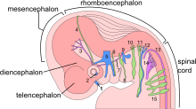
The embryo's nervous system at six weeks.
In the fifth week, the alar plate of the prosencephalon expands to form the cerebral hemispheres (the telencephalon). The basal plate becomes the diencephalon.
The diencephalon, mesencephalon and rhombencephalon constitute the brain stemof the embryo. It continues to flex at the mesencephalon. The rhombencephalon folds posteriorly, which causes its alar plate to flare and form the fourth ventricle of the brain. The pons and the cerebellum form in the upper part of the rhombencephalon, whilst the medulla oblongata forms in the lower part.

The embryo's brain at four weeks.
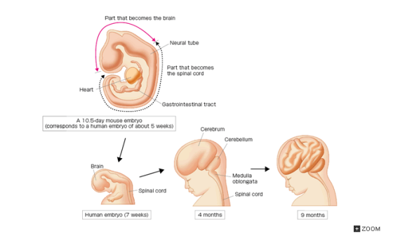

The part of the skull where the brain sits is called the cranium. The base, or lower part, of the brain is connected to the spinal cord. Together, the brain and spinal cord are known as the central nervous system (CNS). Many nerves send electrical signals to and from the brain and spinal cord.
Synapses of the mammalian CNS are asymmetric sites of cell-cell adhesion between nerve cells. They are designed to mediate the rapid and efficient transmission of signals from the presynaptic bouton of one neuron to the postsynaptic plasma membrane of a second neuron. Significant progress has been made in the characterization of the structural, functional and developmental assembly of CNS synapses. Recent progress has been made in understanding the molecular and cellular mechanisms that underlie synaptogenesis, in particular, that of glutamatergic synapses of the CNS. The multimeric protein complexes at CNS synapses. In recent years, numerous molecules have been identified that are likely to play fundamental roles in the assembly and maintenance of CNS synapses. A common theme emerging from these studies is that both the presynaptic active zone and PSD are built from several multi-molecular protein complexes.
Add Answer to:
Describe in detail the development of the neural tube, early
brain and spinal cord development in...
How does exposure to androgens (testosterone) early in life masculinize the brain and spinal cord in...
How does exposure to androgens (testosterone) early in life masculinize the brain and spinal cord in rats? What happens to the hypothalamus in males, compared with females?
Describe the progression and stages of embryo development from fertilization to an early embryo that has...
Describe the progression and stages of embryo development from fertilization to an early embryo that has a fully formed neural tube. Include the following terms (at least) in your description: gastrulation, organogenesis (neurulation), zygote, cleavage, blastula, morula. Explain the functional differences of extraembryonic membranes in terrestrial vertebrates.
11. compare the early embryonic spinal cord to the full developed sponal cord 12. ourline the...
11. compare the early embryonic spinal cord to the full developed sponal cord 12. ourline the divisions of the brain and brain stem and the major structures located in each region. 14. in general, describe rhe formation of the PNS, differentiating cranial and sensory nerves as well as para sympathetic and sympathetic. 16 discuss the strutures derives from pharyngeal arches, puches and/or clefts 17. list structures derives from aortic arch 1-6.
A patient with inflammation of the brain and spinal cord has
A patient with inflammation of the brain and spinal cord has
.Primary spinal cord injury involves damage to vertebral or neural tissues from compression, traction, or shearing...
.Primary spinal cord injury involves damage to vertebral or neural tissues from compression, traction, or shearing forces. Secondary spinal cord injury is related to ischemia, excitotoxicity, inflammation, edema, oxidative damage, and activation of necrotic and apoptotic cell death; it begins within minutes after injury and continues for weeks. Please describe an example of both a primary and secondary spinal cord injury. and describe how the patient experienced both primary and secondary injuries. Be sure to describe symptoms, implications, and testing...
Primary spinal cord injury involves damage to vertebral or neural tissues from compression, traction, or shearing...
Primary spinal cord injury involves damage to vertebral or neural tissues from compression, traction, or shearing forces. Secondary spinal cord injury is related to ischemia, excitotoxicity, inflammation, edema, oxidative damage, and activation of necrotic and apoptotic cell death; it begins within minutes after injury and continues for weeks. Please describe an example of both a primary and secondary spinal cord injury. Some learners will use the same injury, such as a fall off of a horse, and describe how the...
A patient with inflammation of the brain and spinal cord has Flashcards
A patient with inflammation of the brain and spinal cord has Flashcards
The human brain and spinal cord are immersed in the cerebrospinal fluid. The fluid is normally...
 The human brain and spinal cord are immersed in the
cerebrospinal fluid. The fluid is normally continuous between the
cranial and spinal cavities and exerts a pressure of 100 to 200 mm
of H2O above the prevailing atmospheric pressure. In
medical work, pressures are often measured in units of mm of
H2O because body fluids, including the cerebrospinal
fluid, typically have nearly the same density as water. The
pressure of the cerebrospinal fluid can be measured by means of a...
The human brain and spinal cord are immersed in the
cerebrospinal fluid. The fluid is normally continuous between the
cranial and spinal cavities and exerts a pressure of 100 to 200 mm
of H2O above the prevailing atmospheric pressure. In
medical work, pressures are often measured in units of mm of
H2O because body fluids, including the cerebrospinal
fluid, typically have nearly the same density as water. The
pressure of the cerebrospinal fluid can be measured by means of a...
The human brain and spinal cord are immersed in the cerebrospinal fluid. The fluid is normally...
 The human brain and spinal cord are immersed in the cerebrospinal fluid. The fluid is normally continuous between the cranial and spinal cavities and exerts a pressure of 100 to 200 mm of H2 above the prevailing atmospheric pressure. In medical work, pressures are often measured in units of mm of H2O because body fluids, including the cerebrospinal fluid, typically have the same density as water. The pressure of the cerebrospinal fluid can be measured by means of a spinal...
 The human brain and spinal cord are immersed in the
cerebrospinal fluid. The fluid is normally continuous between the
cranial and spinal cavities and exerts a pressure of 100 to 200 mm
of H2O above the prevailing atmospheric pressure. In
medical work, pressures are often measured in units of mm of
H2O because body fluids, including the cerebrospinal
fluid, typically have nearly the same density as water. The
pressure of the cerebrospinal fluid can be measured by means of a...
The human brain and spinal cord are immersed in the
cerebrospinal fluid. The fluid is normally continuous between the
cranial and spinal cavities and exerts a pressure of 100 to 200 mm
of H2O above the prevailing atmospheric pressure. In
medical work, pressures are often measured in units of mm of
H2O because body fluids, including the cerebrospinal
fluid, typically have nearly the same density as water. The
pressure of the cerebrospinal fluid can be measured by means of a...
 The human brain and spinal cord are immersed in the cerebrospinal fluid. The fluid is normally continuous between the cranial and spinal cavities and exerts a pressure of 100 to 200 mm of H2 above the prevailing atmospheric pressure. In medical work, pressures are often measured in units of mm of H2O because body fluids, including the cerebrospinal fluid, typically have the same density as water. The pressure of the cerebrospinal fluid can be measured by means of a spinal...
Most questions answered within 3 hours.
-
Where is the error in this code sequence?
String s1 = "Hello";
String s2 = "ello";...
asked 11 months ago -
Financial data for Joel de Paris, Inc., for last year
follow:
Joel de Paris, Inc.
Balance...
asked 11 months ago -
Consider this reaction:
Al2(SO4)3 (aq)+ BaCl3
(aq) Al2Cl6 (aq)- +
3BaSO4(s) . What is the...
asked 11 months ago -
Suppose that Savneet is considering increasing her
recent random sample from 20 car rentals to 40...
asked 11 months ago -
Trucks arrive at an unloading terminal at an average rate of 120
per hour.
Trucks arrive...
asked 11 months ago -
Why are methanol and ethanol completely soluble in water while
octanol is not very little soluble....
asked 11 months ago -
A facilities manager at a university reads in a research report
that the mean amount of...
asked 11 months ago -
When the CuSO4 is rehydrated by adding water to the anhydrous
compound, is this an endothermic...
asked 11 months ago -
A ray of sunlight is passing from diamond into crown glass; the
angle of incidence is...
asked 11 months ago -
A block of mass 0.249 kg is placed on top of a light, vertical
spring of...
asked 11 months ago -
how do the kidneys compensate in the presences of acidosis
a) trigger hyperventilate
b) reserve acid...
asked 11 months ago -
Question 501 pts
The rental rate of capital to the firm increases. Which of the
following...
asked 11 months ago