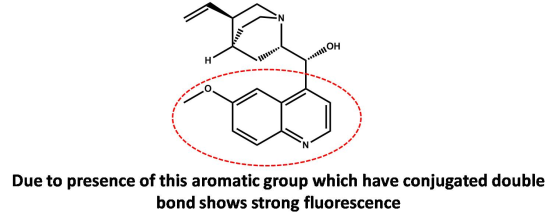
Based on its structure, draw the part of the quinine structure responsible for its strong fluorescence...
Based on its structure, draw the part of the quinine structure responsible for its strong fluorescence and explain your reasoning.
Homework Answers
Add Answer to:
Based on its structure, draw the part of the quinine structure
responsible for its strong fluorescence...
Structure of quinine. Refer to these diagrams wben answering the following questions. a. Which sp...
 please do all the 4 parts neat and clean thanks
structure of quinine. Refer to these diagrams wben answering the following questions. a. Which spectrum (red or blue) is the absorption spectrum? Which spectrum is the fluorescence spectrum? Rationalize your choices. b. What is the best wavelength at which to excite quinine in order to observe its entire fluorescence spectrum? Why? c. What is the best wavelength at which to monitor the fluorescence of quinine-containing solutions? Why? part of the...
please do all the 4 parts neat and clean thanks
structure of quinine. Refer to these diagrams wben answering the following questions. a. Which spectrum (red or blue) is the absorption spectrum? Which spectrum is the fluorescence spectrum? Rationalize your choices. b. What is the best wavelength at which to excite quinine in order to observe its entire fluorescence spectrum? Why? c. What is the best wavelength at which to monitor the fluorescence of quinine-containing solutions? Why? part of the...
Quinine has been used as an anti-malarial drug for many years. Its aqueous solution in certain...
Quinine has been used as an anti-malarial drug for many years. Its aqueous solution in certain acids has been used as a standard in the determination of fluorescence quantum yield of other fluorophores. The structure of quinine is shown below. Quinine in a 1.664-g antimalarial tablet was dissolved in sufficient 0.10 M HCl to give 500 mL of solution. A 10.00 mL aliquot was then diluted to 100.00 mL with the same acid. The fluorescence intensity for the diluted sample...
help me with the highlighted part 3. The fluorescence of each of a series of acidic solutions of quinine was determined five times. The results are given below. Concentration ng mL Fluorescence inten...
 help me with the highlighted part
3. The fluorescence of each of a series of acidic solutions of quinine was determined five times. The results are given below. Concentration ng mL Fluorescence intensity (arbitrary units) 0 4 10 20 30 60 63 60 63 63 40 75 81 79 78 50 104 109 107 101 105 20 21 46 45 4 21 4 Determine the slopes and the ordinate intercepts of the unweighted and weighted regression lines. Calculate, using both...
help me with the highlighted part
3. The fluorescence of each of a series of acidic solutions of quinine was determined five times. The results are given below. Concentration ng mL Fluorescence intensity (arbitrary units) 0 4 10 20 30 60 63 60 63 63 40 75 81 79 78 50 104 109 107 101 105 20 21 46 45 4 21 4 Determine the slopes and the ordinate intercepts of the unweighted and weighted regression lines. Calculate, using both...
Questions in photo. From the Green Synthesis and Study of a Fluorescent Natural Product experiment. Fluorescence Data: Describe the observed under the UV light in the test tube and on the filter...
 Questions in photo. From the Green Synthesis and Study of a
Fluorescent Natural Product experiment.
Fluorescence Data: Describe the observed under the UV light in the test tube and on the filter paper. (e.g. color, brightness etc. fluorescent activity of each test tube of the 4-methylumbelliferone i ) Draw the structure of 4- erone under the neutral, acidic and basic conditions. For the acidic and basic conditions drawa methylumbellif resonance structure to depict a new chromophore likely responsible for its...
Questions in photo. From the Green Synthesis and Study of a
Fluorescent Natural Product experiment.
Fluorescence Data: Describe the observed under the UV light in the test tube and on the filter paper. (e.g. color, brightness etc. fluorescent activity of each test tube of the 4-methylumbelliferone i ) Draw the structure of 4- erone under the neutral, acidic and basic conditions. For the acidic and basic conditions drawa methylumbellif resonance structure to depict a new chromophore likely responsible for its...
Determine the structure based on the molecular formula and its 1H NMR spectrum (and IR spectrum,...
 Determine the structure based on the molecular formula and its
1H NMR spectrum (and IR spectrum, if provided). Show the
work by providing a table summarizing the data from each spectrum
and explain in detail the reasoning. You must analyze each
absorption in the NMR and four absorptions in the IR (if
applicable).
The table for NMR should include: chemical shift (δ
value), the splitting, the number of neighbors, the integration,
and the assignment of the specific protons responsible for the...
Determine the structure based on the molecular formula and its
1H NMR spectrum (and IR spectrum, if provided). Show the
work by providing a table summarizing the data from each spectrum
and explain in detail the reasoning. You must analyze each
absorption in the NMR and four absorptions in the IR (if
applicable).
The table for NMR should include: chemical shift (δ
value), the splitting, the number of neighbors, the integration,
and the assignment of the specific protons responsible for the...
Identify what structure these are based on the NMR and DRAW the structure. 4) Chemical Formula:...
 Identify what structure these are based on the NMR and DRAW the
structure.
4) Chemical Formula: Cl,BrO:S IR: strong broad peak at 3300cm1, strong peak 1720 cm1 Singlet, 3H Quartet, 2H Singlet, 1H Triplet, 2H Triplet, 1H 12 10 PPM
Identify what structure these are based on the NMR and DRAW the
structure.
4) Chemical Formula: Cl,BrO:S IR: strong broad peak at 3300cm1, strong peak 1720 cm1 Singlet, 3H Quartet, 2H Singlet, 1H Triplet, 2H Triplet, 1H 12 10 PPM
Draw the structure of Compound A based on its formula and proton NMR. The integration of...
 Draw the structure of Compound A based on its formula and proton NMR. The integration of the peaks from left to right is: 5:2:1 Compound A MF C_7H_8O Draw the structure of Compound B. Draw the structure of Compound C.
(a) Draw the structure of (4R,5R)-4-ethyl-5-luoro-2-methytheptane. Use solid and dashed wedges to show the proper configurations...
 (a) Draw the structure of (4R,5R)-4-ethyl-5-luoro-2-methytheptane. Use solid and dashed wedges to show the proper configurations of the chiral centers. (0.4 pts) . (b) Now draw a Newman projection for the most stable conformation of the C(4)-C(5) bond. (0.4 pts) (c) Draw a diastereomer of (4R,5R)-4-ethyl-5-fluoro-2-methylheptane (0.2 pts) The specific rotation [a] of pure quinine is-165. A solution containing 9. both quinine and its enantiomer has a specific rotation of -63 Calculate the percentage of quinine present in the mixture?...
One of the following compounds is responsible for the IR spectrum shown below. Draw the structure...
 One of the following compounds is responsible for the IR spectrum shown below. Draw the structure of the responsible compound: 1-butene, 1-butanol, 4-hydroxy-1-butene, methyl propyl ether, or butanoic acid #5. 16
please help in all sections asap!! (1) Draw the structure of Coenzyme A (COA) and explain...
 please help in all sections asap!!
(1) Draw the structure of Coenzyme A (COA) and explain its function based on its structure. (2) Draw the structures of NAD+ & NADH/H+ and explain their function based on their structure. (3) Draw the structures of FAD & FADH2 and explain their function based on their structure.
please help in all sections asap!!
(1) Draw the structure of Coenzyme A (COA) and explain its function based on its structure. (2) Draw the structures of NAD+ & NADH/H+ and explain their function based on their structure. (3) Draw the structures of FAD & FADH2 and explain their function based on their structure.
 please do all the 4 parts neat and clean thanks
structure of quinine. Refer to these diagrams wben answering the following questions. a. Which spectrum (red or blue) is the absorption spectrum? Which spectrum is the fluorescence spectrum? Rationalize your choices. b. What is the best wavelength at which to excite quinine in order to observe its entire fluorescence spectrum? Why? c. What is the best wavelength at which to monitor the fluorescence of quinine-containing solutions? Why? part of the...
please do all the 4 parts neat and clean thanks
structure of quinine. Refer to these diagrams wben answering the following questions. a. Which spectrum (red or blue) is the absorption spectrum? Which spectrum is the fluorescence spectrum? Rationalize your choices. b. What is the best wavelength at which to excite quinine in order to observe its entire fluorescence spectrum? Why? c. What is the best wavelength at which to monitor the fluorescence of quinine-containing solutions? Why? part of the...
 help me with the highlighted part
3. The fluorescence of each of a series of acidic solutions of quinine was determined five times. The results are given below. Concentration ng mL Fluorescence intensity (arbitrary units) 0 4 10 20 30 60 63 60 63 63 40 75 81 79 78 50 104 109 107 101 105 20 21 46 45 4 21 4 Determine the slopes and the ordinate intercepts of the unweighted and weighted regression lines. Calculate, using both...
help me with the highlighted part
3. The fluorescence of each of a series of acidic solutions of quinine was determined five times. The results are given below. Concentration ng mL Fluorescence intensity (arbitrary units) 0 4 10 20 30 60 63 60 63 63 40 75 81 79 78 50 104 109 107 101 105 20 21 46 45 4 21 4 Determine the slopes and the ordinate intercepts of the unweighted and weighted regression lines. Calculate, using both...
 Questions in photo. From the Green Synthesis and Study of a
Fluorescent Natural Product experiment.
Fluorescence Data: Describe the observed under the UV light in the test tube and on the filter paper. (e.g. color, brightness etc. fluorescent activity of each test tube of the 4-methylumbelliferone i ) Draw the structure of 4- erone under the neutral, acidic and basic conditions. For the acidic and basic conditions drawa methylumbellif resonance structure to depict a new chromophore likely responsible for its...
Questions in photo. From the Green Synthesis and Study of a
Fluorescent Natural Product experiment.
Fluorescence Data: Describe the observed under the UV light in the test tube and on the filter paper. (e.g. color, brightness etc. fluorescent activity of each test tube of the 4-methylumbelliferone i ) Draw the structure of 4- erone under the neutral, acidic and basic conditions. For the acidic and basic conditions drawa methylumbellif resonance structure to depict a new chromophore likely responsible for its...
 Determine the structure based on the molecular formula and its
1H NMR spectrum (and IR spectrum, if provided). Show the
work by providing a table summarizing the data from each spectrum
and explain in detail the reasoning. You must analyze each
absorption in the NMR and four absorptions in the IR (if
applicable).
The table for NMR should include: chemical shift (δ
value), the splitting, the number of neighbors, the integration,
and the assignment of the specific protons responsible for the...
Determine the structure based on the molecular formula and its
1H NMR spectrum (and IR spectrum, if provided). Show the
work by providing a table summarizing the data from each spectrum
and explain in detail the reasoning. You must analyze each
absorption in the NMR and four absorptions in the IR (if
applicable).
The table for NMR should include: chemical shift (δ
value), the splitting, the number of neighbors, the integration,
and the assignment of the specific protons responsible for the...
 Identify what structure these are based on the NMR and DRAW the
structure.
4) Chemical Formula: Cl,BrO:S IR: strong broad peak at 3300cm1, strong peak 1720 cm1 Singlet, 3H Quartet, 2H Singlet, 1H Triplet, 2H Triplet, 1H 12 10 PPM
Identify what structure these are based on the NMR and DRAW the
structure.
4) Chemical Formula: Cl,BrO:S IR: strong broad peak at 3300cm1, strong peak 1720 cm1 Singlet, 3H Quartet, 2H Singlet, 1H Triplet, 2H Triplet, 1H 12 10 PPM
 Draw the structure of Compound A based on its formula and proton NMR. The integration of the peaks from left to right is: 5:2:1 Compound A MF C_7H_8O Draw the structure of Compound B. Draw the structure of Compound C.
 (a) Draw the structure of (4R,5R)-4-ethyl-5-luoro-2-methytheptane. Use solid and dashed wedges to show the proper configurations of the chiral centers. (0.4 pts) . (b) Now draw a Newman projection for the most stable conformation of the C(4)-C(5) bond. (0.4 pts) (c) Draw a diastereomer of (4R,5R)-4-ethyl-5-fluoro-2-methylheptane (0.2 pts) The specific rotation [a] of pure quinine is-165. A solution containing 9. both quinine and its enantiomer has a specific rotation of -63 Calculate the percentage of quinine present in the mixture?...
 One of the following compounds is responsible for the IR spectrum shown below. Draw the structure of the responsible compound: 1-butene, 1-butanol, 4-hydroxy-1-butene, methyl propyl ether, or butanoic acid #5. 16
 please help in all sections asap!!
(1) Draw the structure of Coenzyme A (COA) and explain its function based on its structure. (2) Draw the structures of NAD+ & NADH/H+ and explain their function based on their structure. (3) Draw the structures of FAD & FADH2 and explain their function based on their structure.
please help in all sections asap!!
(1) Draw the structure of Coenzyme A (COA) and explain its function based on its structure. (2) Draw the structures of NAD+ & NADH/H+ and explain their function based on their structure. (3) Draw the structures of FAD & FADH2 and explain their function based on their structure.
Most questions answered within 3 hours.
-
Where is the error in this code sequence?
String s1 = "Hello";
String s2 = "ello";...
asked 10 months ago -
Financial data for Joel de Paris, Inc., for last year
follow:
Joel de Paris, Inc.
Balance...
asked 10 months ago -
Consider this reaction:
Al2(SO4)3 (aq)+ BaCl3
(aq) Al2Cl6 (aq)- +
3BaSO4(s) . What is the...
asked 10 months ago -
Suppose that Savneet is considering increasing her
recent random sample from 20 car rentals to 40...
asked 10 months ago -
Trucks arrive at an unloading terminal at an average rate of 120
per hour.
Trucks arrive...
asked 10 months ago -
Why are methanol and ethanol completely soluble in water while
octanol is not very little soluble....
asked 10 months ago -
A facilities manager at a university reads in a research report
that the mean amount of...
asked 10 months ago -
When the CuSO4 is rehydrated by adding water to the anhydrous
compound, is this an endothermic...
asked 10 months ago -
A ray of sunlight is passing from diamond into crown glass; the
angle of incidence is...
asked 10 months ago -
A block of mass 0.249 kg is placed on top of a light, vertical
spring of...
asked 10 months ago -
how do the kidneys compensate in the presences of acidosis
a) trigger hyperventilate
b) reserve acid...
asked 10 months ago -
Question 501 pts
The rental rate of capital to the firm increases. Which of the
following...
asked 10 months ago