what are the mechanisms that clostridum tetani and botulenum, utilize to cause disease in the nervous...
Homework Answers
Botulinum neurotoxins, the most poisonous proteins known to mankind, are a family of seven (types A-G) immunologically distinct proteins synthesized primarily by different strains of the anaerobic bacteria Clostridium botulinum. Botulinum neurotoxin consists of a heavy chain and a light chain linked together by a single disulphide bond. It is synthesised as a relatively inactive single-chain polypeptide with a molecular mass of approximately 150 kDa. It is activated when the polypeptide chain is proteolytically cleaved into the 100-kDa heavy chain and the 50-kDa light chain.

Mechanism of action of botulinum toxin: the light chain of (BT-A) cleaves SNAP-25 (synaptosome-associated protein of 25 KDa) and consequently prevents the release of acetylcholine into the neuromuscular junction.
The clinical syndrome of botulism can occur following ingestion of contaminated food, from colonization of the infant gastrointestinal tract, or from a wound infection.Botulinum toxin (abbreviated either as BTX or BoNT) is broken into 7 neurotoxins (labeled as types A, B, C [C1, C2], D, E, F, and G), which are antigenically and serologically distinct but structurally similar. Human botulism is caused mainly by types A, B, E, and (rarely) F. Although all of these serotypes inhibit acetylcholine release from nerve terminals, their intracellular target proteins, their characteristics of action and their potencies vary substantially. BT type A (BT-A) has been the most widely studied serotype for therapeutic purposes. More recently, BT type B (BT-B) has become commercially available.
Botulinum toxin components

BT Molecular Mode of Action
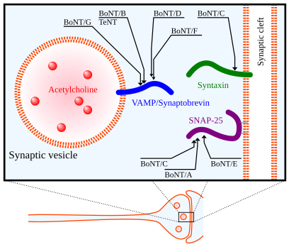
Target molecules of botulinum neurotoxin (abbreviated BoNT) and tetanus neurotoxin (TeNT), toxins acting inside the axon terminal.
When the motoneuron action potential depolarises the axon terminal, acetylcholine is released from the cytosol into the synaptic cleft. This acetylcholine release is performed by a transport protein chain, the soluble N-ethylmaleimide-sensitive factor attachment protein receptor (SNARE) complex. When BT is injected into a target tissue, the heavy chain of the botulinum neurotoxin binds to glycoprotein structures specifically found on cholinergic nerve terminals. This specific docking is the reason for BT’s high selectivity for cholinergic synapses. After internalisation, the light chain of the botulinum neurotoxin binds with high specificity to the SNARE protein complex. The target proteins vary amongst the BT serotypes. BT-A cleaves synaptosome-associated proteins of 25 kDa (SNAP25). BT-B cleaves vesicle-associated membrane protein (VAMP), also known as synaptobrevin II. The light chain’s proteolytic cleavage of the SNARE protein complex prevents the docking of the acetylcholine vesicle on the inner surface of the cellular membrane and results in blockade of vesicle fusion. When the target tissue is a muscle, paresis by chemical denervation occurs. When the target tissue is an exocrine gland, the glandular secretion is blocked. The inhibition of acetylcholine exocytosis by BT is terminated by restoration of the SNARE protein complex turnover. Axonal sprouting and endplate elongation occurs, but is believed to be a transient phenomenon not responsible for the termination of the BT effect.
The seven toxin types (A–G) have different tertiary structures and sequence differences.While the different toxin types all target members of the SNARE family, different toxin types target different SNARE family members. The A, B, and E serotypes cause human botulism, with the activities of types A and B enduring longest in vivo (from several weeks to months).
BT Action on the Central Nervous System
Direct Effects - When BT is injected into a target tissue, it is almost completely bound to the axon terminal. However, when BT-A is applied to treat cervical dystonia, small fractions of the applied BT are distributed systemically and can be detected by an increase in neuromuscular jitter in non-injected muscles. When BT-B is applied to treat cervical dystonia, substantial systemic anticholinergic side-effects can be detected clinically. Despite its systemic distribution, direct BT effects on the central nervous system have not been reported, since botulinum neurotoxin with its size of 150 kDa cannot penetrate the blood-brain barrier. BT, however, could reach the central nervous system by retrograde axonal transport. Indeed, such retrograde axonal transport has been detected for BT with radioactively labelled botulinum neurotoxin. However, the retrograde axonal transport was so slow that the applied BT was likely to be inactivated before it reached the central nervous system. Transsynaptic transport was not observed. BT action upon Renshaw cells was only demonstrated after intraspinal injection.
Indirect Effects - Effects of BT on the neuromuscular synapse and on the muscle spindle organs can produce various indirect effects on the central nervous system. On the spinal level, BT produces reflex inhibition of α-motoneurons by γ-motoneuron blockade and subsequent Ia/II afferent input suppression.In patients with upper limb dystonia, BT can normalise the altered reciprocal inhibition between flexor and extensor muscles. A similar effect was also demonstrated in patients with essential tremor. EMG changes of the contralateral ocular muscles after injection of BT into the lateral rectus muscle also suggest central effects. On the supraspinal level, BT can normalise altered intracortical inhibition and altered somatosensory evoked potentials. Although BT can enhance some aspects of cortical activation, it fails to improve the impaired activation of the primary motor cortex seen in writer’s cramp.
Mechanism of action of Clostridium tetani
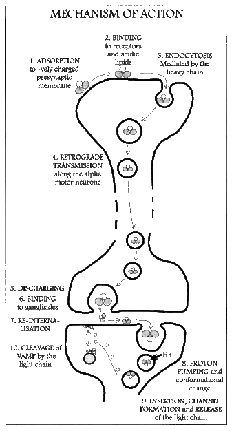
Mechanism of action of tetanospasmin
Tetanus toxin is an extremely potent neurotoxin produced by the vegetative cell of Clostridium tetani in anaerobic conditions, causing tetanus. It has no known function for clostridia in the soil environment where they are normally encountered. It is also called spasmogenic toxin, or TeNT. The LD50 of this toxin has been measured to be approximately 2.5-3 ng/kg making it second only to botulinum toxin (LD50 2 ng/kg) as the deadliest toxin in the world. C. tetani also produces the exotoxin tetanolysin, a hemolysin, that causes destruction of tissues. Tetanus toxin spreads through tissue spaces into the lymphatic and vascular systems. It enters the nervous system at the neuromuscular junctions and migrates through nerve trunks and into the central nervous system (CNS) by retrograde axonal transport by using dyneins.
The tetanus toxin protein has a molecular weight of 150 kDa. It is translated from the tetX gene as one protein which is subsequently cleaved into two parts: a 100 kDa heavy or B-chain and a 50 kDa light or A-chain. The chains are connected by a disulfide bond.
- The B-chain binds to dissialogangliosides (GD2 and GD1b) on the neuronal membrane and contains a translocation domain which aids the movement of the protein across that membrane and into the neuron.
- The A-chain, a zinc endopeptidase, attacks the vesicle-associated membrane protein (VAMP).
The TetX gene encoding this protein is located on the PE88 plasmid.
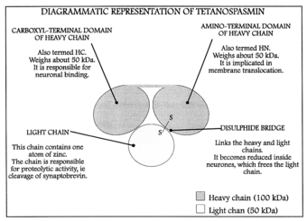
Structure of tetanospasmin
The mechanism of TeNT action can be broken down and discussed in these different steps:
Transport
- Specific binding in the periphery neurons
- Retrograde axonal transport to the CNS inhibitory interneurons
- Transcytosis from the axon into the inhibitory interneurons
Action
- Temperature- and pH-mediated translocation of the light chain into the cytosol
- Reduction of the disulfide bridge to thiols, severing the link between the light and heavy chain
- Cleavage of synaptobrevin
The first three steps outline the travel of tetanus from the peripheral nervous system to where it is taken up to the CNS and has its final effect. The last three steps document the changes necessary for the final mechanism of the neurotoxin.
Transport to the CNS inhibitory interneurons begins with the B-chain mediating the neurospecific binding of TeNT to the nerve terminal membrane. It binds to GT1b polysialogangliosides, similarly to the C. botulinum neurotoxin. It also binds to another poorly characterized GPI-anchored protein receptor more specific to TeNT. Both the ganglioside and the GPI-anchored protein are located in lipid microdomains and both are requisite for specific TeNT binding. Once it is bound, the neurotoxin is then endocytosed into the nerve and begins to travel through the axon to the spinal neurons. The next step, transcytosis from the axon into the CNS inhibitory interneuron, is one of the least understood parts of TeNT action. At least two pathways are involved, one that relies on the recycling of synaptic vesicle 2 (SV2) system and one that does not. Once the vesicle is in the inhibitory interneuron, its translocation is mediated by pH and temperature, specifically a low or acidic pH in the vesicle and standard physiological temperatures. Once the toxin has been translocated into the cytosol, chemical reduction of the disulfide bond to separate thiols occurs, mainly by the enzyme NADPH-thioredoxin reductase-thioredoxin. The light chain is then free to cleave the Gln76-Phe77 bond of synaptobrevin. Cleavage of synaptobrevin affects the stability of the SNARE core by restricting it from entering the low-energy conformation, which is the target for NSF binding. Synaptobrevin is an integral V-SNARE necessary for vesicle fusion to membranes. The final target of TeNT is the cleavage of synaptobrevin and, even in low doses, has the effect of interfering with exocytosis of neurotransmitters from inhibitory interneurons. The blockage of the neurotransmitters γ-aminobutyric acid (GABA) and glycine is the direct cause of the physiological effects that TeNT induces. GABA inhibits motor neurons, so by blocking GABA, tetanus toxin causes violent spastic paralysis. The action of the A-chain also stops the affected neurons from releasing excitatory transmitters, by degrading the protein synaptobrevin 2. The combined consequence is dangerous overactivity in the muscles from the smallest sensory stimuli, as the damping of motor reflexes is inhibited, leading to generalized contractions of the agonist and antagonist musculature, termed a "tetanic spasm".
Add Answer to:
what are the mechanisms that clostridum tetani and botulenum,
utilize to cause disease in the nervous...
what are the mechanisms that streptococcus pneumoniae utilize to cause disease in the nervous system?
what are the mechanisms that streptococcus pneumoniae utilize to cause disease in the nervous system?
what ate the mechanisms that pseudomonas aeruginosa utilize to cause disease on skin and in the...
what ate the mechanisms that pseudomonas aeruginosa utilize to cause disease on skin and in the eyes
Please help 1. What is a Coronary artery disease . what is sign and symptom, cause,...
Please help 1. What is a Coronary artery disease . what is sign and symptom, cause, 2. what is Hypertension what is sign and symptom, cause, 3. what is Peripheral arterial disease what is sign and symptom, cause,
Can someone help me i have to write an Final Essays about this paper. 1. Please...
Can someone help me i have to write an Final Essays about this paper. 1. Please discuss the mechanisms that Clostridium tetani and botulinum, Streptococcus pneumonia and Trypanosome cruz and bruce. utilize to cause disease in the nervous system. You must state what disease these microorganisms cause . in addition, please discuss that host immune defenses that the microbes would have to overcome to be success fuel at causing disease (hint:) A. include the three routes of entry to gain...
Please help to answer these question What is the congestive heart failure. What is cause ....
Please help to answer these question What is the congestive heart failure. What is cause . What is left and right heart failure 1. What is a Coronary artery disease . what is sign and symptom, cause, 2. what is Hypertension what is sign and symptom, cause, 3. what is Peripheral arterial disease what is sign and symptom, cause,
I have to write essay or discuss the mechanisms that streptococcus mutans, helicobacter pylori, e.colli and...
I have to write essay or discuss the mechanisms that streptococcus mutans, helicobacter pylori, e.colli and staphylococcus aureus and taenia solium utilize to cause disease in the digestive tract. in addition, list and explain the host immune defenses that microbes would have to overcome to be successful at causing disease.
What is Alzhemier's Disease? What is the cause, symptoms and treatments for Alzhemier's Disease?
What is Alzhemier's Disease? What is the cause, symptoms and treatments for Alzhemier's Disease?
D, and D2 cause sympton A with probabilities 04 and 0.8 respectively. Suppose 6% of a...
 D, and D2 cause sympton A with probabilities 04 and 0.8 respectively. Suppose 6% of a population have disease Di and 2% have disease Da. Assume that the only possible causes of symptom A are D and D2, and that no one carries more than one of those two diseases. (a) What percent of the population have symptom A? (b) Let a randomly selected person from the population have symptom A. What is the chance be/sbe carries disease Di?
what is/are the cause/causes of the disease breast cancer
what is/are the cause/causes of the disease breast cancer
List the four central nervous system malformations that cause polyhydramnios.
List the four central nervous system malformations that cause polyhydramnios.
 D, and D2 cause sympton A with probabilities 04 and 0.8 respectively. Suppose 6% of a population have disease Di and 2% have disease Da. Assume that the only possible causes of symptom A are D and D2, and that no one carries more than one of those two diseases. (a) What percent of the population have symptom A? (b) Let a randomly selected person from the population have symptom A. What is the chance be/sbe carries disease Di?
Most questions answered within 3 hours.
-
Where is the error in this code sequence?
String s1 = "Hello";
String s2 = "ello";...
asked 11 months ago -
Financial data for Joel de Paris, Inc., for last year
follow:
Joel de Paris, Inc.
Balance...
asked 11 months ago -
Consider this reaction:
Al2(SO4)3 (aq)+ BaCl3
(aq) Al2Cl6 (aq)- +
3BaSO4(s) . What is the...
asked 11 months ago -
Suppose that Savneet is considering increasing her
recent random sample from 20 car rentals to 40...
asked 11 months ago -
Trucks arrive at an unloading terminal at an average rate of 120
per hour.
Trucks arrive...
asked 11 months ago -
Why are methanol and ethanol completely soluble in water while
octanol is not very little soluble....
asked 11 months ago -
A facilities manager at a university reads in a research report
that the mean amount of...
asked 11 months ago -
When the CuSO4 is rehydrated by adding water to the anhydrous
compound, is this an endothermic...
asked 11 months ago -
A ray of sunlight is passing from diamond into crown glass; the
angle of incidence is...
asked 11 months ago -
A block of mass 0.249 kg is placed on top of a light, vertical
spring of...
asked 11 months ago -
how do the kidneys compensate in the presences of acidosis
a) trigger hyperventilate
b) reserve acid...
asked 11 months ago -
Question 501 pts
The rental rate of capital to the firm increases. Which of the
following...
asked 11 months ago