if you to monitor the activity of any of the serine proteases as they degrade protein,...
if you to monitor the activity of any of the serine proteases as they degrade protein, you would note that the serine protease does not cut all of the recognition sites in the substratebat once- the recognition sites in the surface of the subrate protein are the first to be cut. Explain
Homework Answers
Answer:
Pancreatic exocrine secretions- enzymes: serine proteases (trypsinogen, chymotrypsinogen, subtilinases), These are inactive and become active in the duodenum and later involve in breaking down the polymer macro molecules such as proteins into simple monomers as amino acids by "hydrolysis".
The crucial component in the mechanism of serine protease enzyme catalysis as they bind with the substrate is the formation of catalytic traid (His 57, ser 195 and aspar 102). This catalytic traid have the capability to recognize the catalytic sites of the substrate polypeptide as these substrates are in complex tertiary structure forms due to folding. The significant serine proteases geometry is enable them to recognize the substrate cleavage sites and they characteristically bind at the transition state and finally declines the overall activation energy of the reaction and thereby enables the significant catalytic ability of the enzyme. These zymozens (serine proteases) are initially inactive in the pancreas. These are inactive precursors of the serine proteases synthesized in the pancreas, incase if they become active where they synthesized without recognizing the substrate site may leading to autolysis (self-digestion) of the enzyme. Further this self digestion due to premature activation of serine proteases result in acute pancreatitis.
Activation of the proteases is prerequisite during the catalysis of the substrate polypeptide. Hydroxyl group of serine proteases acts as nucleophile and the substrate polypeptide will be exposed with active site thereby a scissile covalent bond is mixed with the enzyme active site result in the proteolysis.
Add Answer to:
if you to monitor the activity of any of the serine
proteases as they degrade protein,...
1) (10 pts) Serine proteases are enzymes that cleave peptide bonds in proteins. Explain using words...
 1) (10 pts) Serine proteases are enzymes that cleave peptide bonds in proteins. Explain using words (drawings OR both) why serine proteases cleave either before or after different amino acid residues. Talk about at least two of the following proteases: Chymotrypsin, Trypsin, or Elastase. mod 2) (10pts) Below is a hypothetical peptide sequence. Give the peptide fragments that will occur by enzymatic degradation using Trypsin and Chymotrypsin DARSKWKSENLIRTY 3) (10 pts) All superfamilies of serine proteases use the catalytic triad...
1.. Example questions to guide you through the mechanism study of Serine proteases Describe substrate binding,...
1.. Example questions to guide you through the mechanism study of Serine proteases Describe substrate binding, including the role and chemical nature of the "specificity pocket" in chymotrypsin, and which peptide bond in the substrate (relative to the specificity group) will be cleaved. Draw the structure of the catalytic triad at the beginning of the reaction, and explain how the states of ionization and hydrogen bonding pattern of those 3 groups change step by step during catalysis. Explain the role...
An mRNA is translated into a protein in a cell-free extract in the absence of presence...
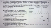 An mRNA is translated into a protein in a cell-free extract in the absence of presence of microsomes or endoplasmic reticulum. Samples are treated as follows: (I) no treatment, (II) addition of protease, (IIl) addition of protease and detergent, and (IV) disruption of mircosomes and addition of endoH, an enzyme removing N-linked sugars that are added in the ER. Analysis by gel electrophoresis is shown in the figure. 7. MICROSOMESİ MICROSOMES ABSENT PRESENT TREATMENT protease a. Proteih synthesis at the...
An F' plasmid from an exotic marine bacterium that can degrade petroleum when mated with an...
An F' plasmid from an exotic marine bacterium that can degrade petroleum when mated with an F-E. coli that cannot degrade petroleum results in an E. coli F' that can degrade petroleum. After sequencing the F' plasmid and comparing it to the F sequence, you see that it has about 6000 base pairs of additional DNA. You suspect that there is a gene in that sequence that encodes a protein that degrades petroleum. Describe how you would use reverse genetics...
Kinetics nndInbibition 3. Based on your understanding of this experiment, propose a rationale for why skim...
 Kinetics nndInbibition 3. Based on your understanding of this experiment, propose a rationale for why skim milk stays fresher longer than whole milk? What pattern did you see with the different juice sources that were used? Rank all three in order of protease activity, pH, and clearing diameter. Do any patterns emerge? 4. 5. Why do the pH and protease rankings differ between fresh fruits but are similar for cooked fruits? 6. Knowing proteases alter the appearance of the milk,...
In-Class Activity #12, Protein Tratlicking Worksheet 1. You are interested in four different proteins in a...
 In-Class Activity #12, Protein Tratlicking Worksheet 1. You are interested in four different proteins in a yeast cell: • protein 1 is a cytosolic protein protein 2 is a secreted protein • protein 3 is a nuclear protein protein 4 is a cell surface membrane protein with N-terminal end in extra-cellular space. a. You plan to study how the proteins are localized to their specific destination by creating the following mutations in the genes encoding proteins 1-4. Indicate how the...
O ACTIVITY 5.4.1 Synthesis of a Protein: A Simulation Activity In this activity, you will be...
 O ACTIVITY 5.4.1 Synthesis of a Protein: A Simulation Activity In this activity, you will be provided with the DNA nucleotide sequence that codes for a hypothetical protein. The code will be provided to you in three fragments. You will have to tran- scribe the code into mRNA, remove an intron segment, and translate the mRNA into the protein. In addition, you will have to identify the beginning fragment the middle fragment, and the end fragment. Sequence A TCTTCCCTCCTAAACGTTCAACCGGTTCTTAATCCGC CGCCAGGGCCCCGCCCCTCAGAAGTTGGT...
You include an innhibitor of the protein kinase activity of TFIIH in an in virto transcription....
You include an innhibitor of the protein kinase activity of TFIIH in an in virto transcription. What step in transcription would you expect to see blocked? Decribe an experiment you would run to test your hypothesis? Please explain the experiment part to the best of your ability.
Any help would be appreciated! S y of a protease activity of Science you decided to...
 Any help would be appreciated!
S y of a protease activity of Science you decided to develop a series of peptide sequence to probe sequence versus activity profiles. boa experimenting the lo n g e sequence versus activity profiles You deseo DNA plecamid sequence entire plasmid not shown-just the ce desired Answer the e plasmid not show-just the site of interest and DNA questions using these sequences (24 pts) Plasmid Sequence (selected insert portion)" TCGAGCCGATATCCTGCAGCGATGAAGCTTGOTAGOGTCGACTAACACTGCAGOTCOACCO AGCGCTATAGGACOTCGCTACTTCGAACCATCGCAGCTGATTGTGACGTCCAGAR rege Althe t he...
Any help would be appreciated!
S y of a protease activity of Science you decided to develop a series of peptide sequence to probe sequence versus activity profiles. boa experimenting the lo n g e sequence versus activity profiles You deseo DNA plecamid sequence entire plasmid not shown-just the ce desired Answer the e plasmid not show-just the site of interest and DNA questions using these sequences (24 pts) Plasmid Sequence (selected insert portion)" TCGAGCCGATATCCTGCAGCGATGAAGCTTGOTAGOGTCGACTAACACTGCAGOTCOACCO AGCGCTATAGGACOTCGCTACTTCGAACCATCGCAGCTGATTGTGACGTCCAGAR rege Althe t he...
Biochem- please answer all You need to denature a heterodimeric protein that uses both ionic interactions...
 Biochem- please answer all
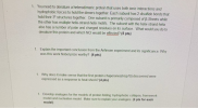
You need to denature a heterodimeric protein that uses both ionic interactions and hydrophobic forces to hold the dimers together. Each subunit has 2 disulfide bonds that hold their 3 structures together. One subunit is primarily composed of P-Sheets while the other has multiple helix-strand-helix motifs. The subunit with the helix-strand-helix also has a number of polar and charged residues on its surface. What would you do to denature this protein and which NCI would be...
Biochem- please answer all
You need to denature a heterodimeric protein that uses both ionic interactions and hydrophobic forces to hold the dimers together. Each subunit has 2 disulfide bonds that hold their 3 structures together. One subunit is primarily composed of P-Sheets while the other has multiple helix-strand-helix motifs. The subunit with the helix-strand-helix also has a number of polar and charged residues on its surface. What would you do to denature this protein and which NCI would be...
 1) (10 pts) Serine proteases are enzymes that cleave peptide bonds in proteins. Explain using words (drawings OR both) why serine proteases cleave either before or after different amino acid residues. Talk about at least two of the following proteases: Chymotrypsin, Trypsin, or Elastase. mod 2) (10pts) Below is a hypothetical peptide sequence. Give the peptide fragments that will occur by enzymatic degradation using Trypsin and Chymotrypsin DARSKWKSENLIRTY 3) (10 pts) All superfamilies of serine proteases use the catalytic triad...
 An mRNA is translated into a protein in a cell-free extract in the absence of presence of microsomes or endoplasmic reticulum. Samples are treated as follows: (I) no treatment, (II) addition of protease, (IIl) addition of protease and detergent, and (IV) disruption of mircosomes and addition of endoH, an enzyme removing N-linked sugars that are added in the ER. Analysis by gel electrophoresis is shown in the figure. 7. MICROSOMESİ MICROSOMES ABSENT PRESENT TREATMENT protease a. Proteih synthesis at the...
 Kinetics nndInbibition 3. Based on your understanding of this experiment, propose a rationale for why skim milk stays fresher longer than whole milk? What pattern did you see with the different juice sources that were used? Rank all three in order of protease activity, pH, and clearing diameter. Do any patterns emerge? 4. 5. Why do the pH and protease rankings differ between fresh fruits but are similar for cooked fruits? 6. Knowing proteases alter the appearance of the milk,...
 In-Class Activity #12, Protein Tratlicking Worksheet 1. You are interested in four different proteins in a yeast cell: • protein 1 is a cytosolic protein protein 2 is a secreted protein • protein 3 is a nuclear protein protein 4 is a cell surface membrane protein with N-terminal end in extra-cellular space. a. You plan to study how the proteins are localized to their specific destination by creating the following mutations in the genes encoding proteins 1-4. Indicate how the...
 O ACTIVITY 5.4.1 Synthesis of a Protein: A Simulation Activity In this activity, you will be provided with the DNA nucleotide sequence that codes for a hypothetical protein. The code will be provided to you in three fragments. You will have to tran- scribe the code into mRNA, remove an intron segment, and translate the mRNA into the protein. In addition, you will have to identify the beginning fragment the middle fragment, and the end fragment. Sequence A TCTTCCCTCCTAAACGTTCAACCGGTTCTTAATCCGC CGCCAGGGCCCCGCCCCTCAGAAGTTGGT...
 Any help would be appreciated!
S y of a protease activity of Science you decided to develop a series of peptide sequence to probe sequence versus activity profiles. boa experimenting the lo n g e sequence versus activity profiles You deseo DNA plecamid sequence entire plasmid not shown-just the ce desired Answer the e plasmid not show-just the site of interest and DNA questions using these sequences (24 pts) Plasmid Sequence (selected insert portion)" TCGAGCCGATATCCTGCAGCGATGAAGCTTGOTAGOGTCGACTAACACTGCAGOTCOACCO AGCGCTATAGGACOTCGCTACTTCGAACCATCGCAGCTGATTGTGACGTCCAGAR rege Althe t he...
Any help would be appreciated!
S y of a protease activity of Science you decided to develop a series of peptide sequence to probe sequence versus activity profiles. boa experimenting the lo n g e sequence versus activity profiles You deseo DNA plecamid sequence entire plasmid not shown-just the ce desired Answer the e plasmid not show-just the site of interest and DNA questions using these sequences (24 pts) Plasmid Sequence (selected insert portion)" TCGAGCCGATATCCTGCAGCGATGAAGCTTGOTAGOGTCGACTAACACTGCAGOTCOACCO AGCGCTATAGGACOTCGCTACTTCGAACCATCGCAGCTGATTGTGACGTCCAGAR rege Althe t he...
 Biochem- please answer all
You need to denature a heterodimeric protein that uses both ionic interactions and hydrophobic forces to hold the dimers together. Each subunit has 2 disulfide bonds that hold their 3 structures together. One subunit is primarily composed of P-Sheets while the other has multiple helix-strand-helix motifs. The subunit with the helix-strand-helix also has a number of polar and charged residues on its surface. What would you do to denature this protein and which NCI would be...
Biochem- please answer all
You need to denature a heterodimeric protein that uses both ionic interactions and hydrophobic forces to hold the dimers together. Each subunit has 2 disulfide bonds that hold their 3 structures together. One subunit is primarily composed of P-Sheets while the other has multiple helix-strand-helix motifs. The subunit with the helix-strand-helix also has a number of polar and charged residues on its surface. What would you do to denature this protein and which NCI would be...
Most questions answered within 3 hours.
-
Where is the error in this code sequence?
String s1 = "Hello";
String s2 = "ello";...
asked 11 months ago -
Financial data for Joel de Paris, Inc., for last year
follow:
Joel de Paris, Inc.
Balance...
asked 11 months ago -
Consider this reaction:
Al2(SO4)3 (aq)+ BaCl3
(aq) Al2Cl6 (aq)- +
3BaSO4(s) . What is the...
asked 11 months ago -
Suppose that Savneet is considering increasing her
recent random sample from 20 car rentals to 40...
asked 11 months ago -
Trucks arrive at an unloading terminal at an average rate of 120
per hour.
Trucks arrive...
asked 11 months ago -
Why are methanol and ethanol completely soluble in water while
octanol is not very little soluble....
asked 11 months ago -
A facilities manager at a university reads in a research report
that the mean amount of...
asked 11 months ago -
When the CuSO4 is rehydrated by adding water to the anhydrous
compound, is this an endothermic...
asked 11 months ago -
A ray of sunlight is passing from diamond into crown glass; the
angle of incidence is...
asked 11 months ago -
A block of mass 0.249 kg is placed on top of a light, vertical
spring of...
asked 11 months ago -
how do the kidneys compensate in the presences of acidosis
a) trigger hyperventilate
b) reserve acid...
asked 11 months ago -
Question 501 pts
The rental rate of capital to the firm increases. Which of the
following...
asked 11 months ago