
Homework Answers
A: Please go through following schematics

B Restriction map
Add Answer to:
B4. Answer ALL parts: The diagram below shows the restriction pattern of a plasmid cut with...
34. Answer ALL parts: The diagram below shows the restriction pattern of a plasmid cut with...
 34. Answer ALL parts: The diagram below shows the restriction pattern of a plasmid cut with the enzymes Smal, Bglil and Aval. The digests are carried out with each enzyme alone and then with different combinations of the three enzymes. Enzyme Fragment sizes kb Smal 5.9 4.3 BgII 8.2 2.0 Aval 5.3 4.9 Smal+ BglII 5.4 2.8 0.5 Smal+ Aval 3.3 2.6 2.3 2.0 Smal+-Bg!!! 5.3 2.1 2.0 0.8 (a) Draw an agarose gel with restriction fragments from the data...
Restriction Mapping Below is a restriction map for the plasmid PGEN101 (total length - 20 kb)....
 Restriction Mapping Below is a restriction map for the plasmid PGEN101 (total length - 20 kb). Using this map as a guide, give the number of restriction fragments along with their associated lengths that would result from digesting PGEN101 with the restriction enzymes EcoRI, BamHII, anda combination of EcoRI + BamHI. BamHI BamHI BamHI / PGEN101 (20 kb) Mb EcoRI Digest Performed Size Emments Obtained EcoRI........ BamHI.. EcoRI + BamHI.... Two freshmen college students, interested in becoming gene jocks, performed...
Module 2 homework Laurel Jacqmai 0341 - Spring 20-MEEK > Activities and Due Dates > Module...
 Module 2 homework Laurel Jacqmai 0341 - Spring 20-MEEK > Activities and Due Dates > Module 2 homework Assignment Score: 20% Resources Hint Check Ans < Question 2 of 5 > EcoRI Control EcoRI BamHI BamHI Base pairs 10 kb Samples of a plasmid containing a segment of unknown DNA are digested using the restriction enzymes EcoRI, BamHI, and a combination of EcoRI and BamHI. The digests are then run on an agarose gel in order to separate the resulting...
Chromosomal and plasmid DNA can be cut into manageable pieces by restriction enzymes. Using agarose gel...
 Chromosomal and plasmid DNA can be cut into manageable pieces by
restriction enzymes. Using agarose gel electrophoresis, the DNA
fragments can be separated on a gel, based on their lengths. In
order to see the fragments, a stain is typically added to the gel.
The size of each fragment can be determined by comparing each one
to a DNA molecular weight marker of known size.
Below is a map of pBR22 plasmid. The position and base pair
number of the...
Chromosomal and plasmid DNA can be cut into manageable pieces by
restriction enzymes. Using agarose gel electrophoresis, the DNA
fragments can be separated on a gel, based on their lengths. In
order to see the fragments, a stain is typically added to the gel.
The size of each fragment can be determined by comparing each one
to a DNA molecular weight marker of known size.
Below is a map of pBR22 plasmid. The position and base pair
number of the...
The table below presents the results of different digests of a plasmid. All sizes are in...
The table below presents the results of different digests of a plasmid. All sizes are in base pairs. Digest Size of Fragments HindIII 4.00 kb BamHI 2.35 kb, 1.65 kb EcoRI 4.00 kb, 2.50 kb,1.50 kb HindIII + BamHI 1.65 kb, 1.50 kb, 0.85 kb HindIII + EcoRI 3.50 kb, 3.00 kb, 2.50kb, 1.00 kb, 0.50 kb BamHI + EcoRI 2.00kb, 1.85kb, 1.65kb, 1.5kb, 0.35kb, 0.50 kb One of the enzymes used only partially digested the DNA. Which enzyme is...
You have determined that a bacterial strain you are working with contains a single type of...
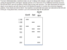 You have determined that a bacterial strain you are working with
contains a single type of plasmid. You isolate the plasmid DNA and
digest separate portions of it with each of two different
restriction enzymes, BamHI and HpaI, and also perform a double
digest using both enzymes. You then fractionate the enzyme digests
on an agarose gel and stain the gel with ethidium bromideto
visualize the restriction fragment patterns. Your results are shown
below. Size markers (in nucleotide base pairs)...
You have determined that a bacterial strain you are working with
contains a single type of plasmid. You isolate the plasmid DNA and
digest separate portions of it with each of two different
restriction enzymes, BamHI and HpaI, and also perform a double
digest using both enzymes. You then fractionate the enzyme digests
on an agarose gel and stain the gel with ethidium bromideto
visualize the restriction fragment patterns. Your results are shown
below. Size markers (in nucleotide base pairs)...
Calculate the restriction map of plasmid JLB51given the following restriction digest data. HindIII 3.82 0.18 kb...
 Calculate the restriction map of plasmid JLB51given the following restriction digest data. HindIII 3.82 0.18 kb BamHI 2.35, 1.65 kb EcoRI 3.00, 1.0 kb HindIII, BamHI 2.35, 1.2, 0.27, 0.18 kb HindIII, EcoRI 1.87, 1.00, 0.95, 0.18 kb BamHI, EcoRI 1.6, 1.4, 0.75, 0.25 kb draw the circular map
MICROBIOLOGY HELP Exercise: Understanding restriction enzyme digests 1. Below is an image of a plasmid. Various...
 MICROBIOLOGY HELP
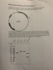
Exercise: Understanding restriction enzyme digests 1. Below is an image of a plasmid. Various parts of the pl location of two restriction enzyme sites fiction enzyme sites (EcoRI and Hindill) are shown. id. Various parts of the plasmid as well as the ECOR PYFG drugR A researcher performs some restriction enzyme digests of this plasmid. In separate tubes, he digests the plasmid with EcoRI alone. HindIll alone, and EcoRI and Hindill together. He also sets up a sample...
MICROBIOLOGY HELP
Exercise: Understanding restriction enzyme digests 1. Below is an image of a plasmid. Various parts of the pl location of two restriction enzyme sites fiction enzyme sites (EcoRI and Hindill) are shown. id. Various parts of the plasmid as well as the ECOR PYFG drugR A researcher performs some restriction enzyme digests of this plasmid. In separate tubes, he digests the plasmid with EcoRI alone. HindIll alone, and EcoRI and Hindill together. He also sets up a sample...
9. On Worksheet 16.IIIB is a restriction map of bacteriophage lambda. You digest some lambda DNA...
 9. On Worksheet 16.IIIB is a restriction map of bacteriophage lambda. You digest some lambda DNA with the enzymes BamHI and HindIII separately and then load the fragments into an agarose gel and perform electrophoresis. Next, you perform a Southern analysis using the 4,878-bp EcoRI lambda fragment as a probe. a. Draw a picture of the electrophoresis gel, using the outline of the stained electrophoresis gel in Worksheet 16.IIIB (the two smallest HindIII fragments will run off the gel.) b....
please help me with these questions Lab 8 Extension Activity: Plasmid Mapping and Restriction Enzymes Mapping...
 please help me with these questions
Lab 8 Extension Activity: Plasmid Mapping and Restriction Enzymes Mapping the Plasmid The first step in mapping a plasmid is to determine how many times a restriction site is found on that plasmid. Examine the results for plasmid 55 as an example. The data given in the following table are for the double digest using EcoRI and Pstl. Also, giving are the data for single digests by the individual enzymes. The numbers in the...
please help me with these questions
Lab 8 Extension Activity: Plasmid Mapping and Restriction Enzymes Mapping the Plasmid The first step in mapping a plasmid is to determine how many times a restriction site is found on that plasmid. Examine the results for plasmid 55 as an example. The data given in the following table are for the double digest using EcoRI and Pstl. Also, giving are the data for single digests by the individual enzymes. The numbers in the...
 34. Answer ALL parts: The diagram below shows the restriction pattern of a plasmid cut with the enzymes Smal, Bglil and Aval. The digests are carried out with each enzyme alone and then with different combinations of the three enzymes. Enzyme Fragment sizes kb Smal 5.9 4.3 BgII 8.2 2.0 Aval 5.3 4.9 Smal+ BglII 5.4 2.8 0.5 Smal+ Aval 3.3 2.6 2.3 2.0 Smal+-Bg!!! 5.3 2.1 2.0 0.8 (a) Draw an agarose gel with restriction fragments from the data...
 Restriction Mapping Below is a restriction map for the plasmid PGEN101 (total length - 20 kb). Using this map as a guide, give the number of restriction fragments along with their associated lengths that would result from digesting PGEN101 with the restriction enzymes EcoRI, BamHII, anda combination of EcoRI + BamHI. BamHI BamHI BamHI / PGEN101 (20 kb) Mb EcoRI Digest Performed Size Emments Obtained EcoRI........ BamHI.. EcoRI + BamHI.... Two freshmen college students, interested in becoming gene jocks, performed...
 Module 2 homework Laurel Jacqmai 0341 - Spring 20-MEEK > Activities and Due Dates > Module 2 homework Assignment Score: 20% Resources Hint Check Ans < Question 2 of 5 > EcoRI Control EcoRI BamHI BamHI Base pairs 10 kb Samples of a plasmid containing a segment of unknown DNA are digested using the restriction enzymes EcoRI, BamHI, and a combination of EcoRI and BamHI. The digests are then run on an agarose gel in order to separate the resulting...
 Chromosomal and plasmid DNA can be cut into manageable pieces by
restriction enzymes. Using agarose gel electrophoresis, the DNA
fragments can be separated on a gel, based on their lengths. In
order to see the fragments, a stain is typically added to the gel.
The size of each fragment can be determined by comparing each one
to a DNA molecular weight marker of known size.
Below is a map of pBR22 plasmid. The position and base pair
number of the...
Chromosomal and plasmid DNA can be cut into manageable pieces by
restriction enzymes. Using agarose gel electrophoresis, the DNA
fragments can be separated on a gel, based on their lengths. In
order to see the fragments, a stain is typically added to the gel.
The size of each fragment can be determined by comparing each one
to a DNA molecular weight marker of known size.
Below is a map of pBR22 plasmid. The position and base pair
number of the...
 You have determined that a bacterial strain you are working with
contains a single type of plasmid. You isolate the plasmid DNA and
digest separate portions of it with each of two different
restriction enzymes, BamHI and HpaI, and also perform a double
digest using both enzymes. You then fractionate the enzyme digests
on an agarose gel and stain the gel with ethidium bromideto
visualize the restriction fragment patterns. Your results are shown
below. Size markers (in nucleotide base pairs)...
You have determined that a bacterial strain you are working with
contains a single type of plasmid. You isolate the plasmid DNA and
digest separate portions of it with each of two different
restriction enzymes, BamHI and HpaI, and also perform a double
digest using both enzymes. You then fractionate the enzyme digests
on an agarose gel and stain the gel with ethidium bromideto
visualize the restriction fragment patterns. Your results are shown
below. Size markers (in nucleotide base pairs)...
 Calculate the restriction map of plasmid JLB51given the following restriction digest data. HindIII 3.82 0.18 kb BamHI 2.35, 1.65 kb EcoRI 3.00, 1.0 kb HindIII, BamHI 2.35, 1.2, 0.27, 0.18 kb HindIII, EcoRI 1.87, 1.00, 0.95, 0.18 kb BamHI, EcoRI 1.6, 1.4, 0.75, 0.25 kb draw the circular map
 MICROBIOLOGY HELP
Exercise: Understanding restriction enzyme digests 1. Below is an image of a plasmid. Various parts of the pl location of two restriction enzyme sites fiction enzyme sites (EcoRI and Hindill) are shown. id. Various parts of the plasmid as well as the ECOR PYFG drugR A researcher performs some restriction enzyme digests of this plasmid. In separate tubes, he digests the plasmid with EcoRI alone. HindIll alone, and EcoRI and Hindill together. He also sets up a sample...
MICROBIOLOGY HELP
Exercise: Understanding restriction enzyme digests 1. Below is an image of a plasmid. Various parts of the pl location of two restriction enzyme sites fiction enzyme sites (EcoRI and Hindill) are shown. id. Various parts of the plasmid as well as the ECOR PYFG drugR A researcher performs some restriction enzyme digests of this plasmid. In separate tubes, he digests the plasmid with EcoRI alone. HindIll alone, and EcoRI and Hindill together. He also sets up a sample...
 9. On Worksheet 16.IIIB is a restriction map of bacteriophage lambda. You digest some lambda DNA with the enzymes BamHI and HindIII separately and then load the fragments into an agarose gel and perform electrophoresis. Next, you perform a Southern analysis using the 4,878-bp EcoRI lambda fragment as a probe. a. Draw a picture of the electrophoresis gel, using the outline of the stained electrophoresis gel in Worksheet 16.IIIB (the two smallest HindIII fragments will run off the gel.) b....
 please help me with these questions
Lab 8 Extension Activity: Plasmid Mapping and Restriction Enzymes Mapping the Plasmid The first step in mapping a plasmid is to determine how many times a restriction site is found on that plasmid. Examine the results for plasmid 55 as an example. The data given in the following table are for the double digest using EcoRI and Pstl. Also, giving are the data for single digests by the individual enzymes. The numbers in the...
please help me with these questions
Lab 8 Extension Activity: Plasmid Mapping and Restriction Enzymes Mapping the Plasmid The first step in mapping a plasmid is to determine how many times a restriction site is found on that plasmid. Examine the results for plasmid 55 as an example. The data given in the following table are for the double digest using EcoRI and Pstl. Also, giving are the data for single digests by the individual enzymes. The numbers in the...
Most questions answered within 3 hours.
-
Where is the error in this code sequence?
String s1 = "Hello";
String s2 = "ello";...
asked 11 months ago -
Financial data for Joel de Paris, Inc., for last year
follow:
Joel de Paris, Inc.
Balance...
asked 11 months ago -
Consider this reaction:
Al2(SO4)3 (aq)+ BaCl3
(aq) Al2Cl6 (aq)- +
3BaSO4(s) . What is the...
asked 11 months ago -
Suppose that Savneet is considering increasing her
recent random sample from 20 car rentals to 40...
asked 11 months ago -
Trucks arrive at an unloading terminal at an average rate of 120
per hour.
Trucks arrive...
asked 11 months ago -
Why are methanol and ethanol completely soluble in water while
octanol is not very little soluble....
asked 11 months ago -
A facilities manager at a university reads in a research report
that the mean amount of...
asked 11 months ago -
When the CuSO4 is rehydrated by adding water to the anhydrous
compound, is this an endothermic...
asked 11 months ago -
A ray of sunlight is passing from diamond into crown glass; the
angle of incidence is...
asked 11 months ago -
A block of mass 0.249 kg is placed on top of a light, vertical
spring of...
asked 11 months ago -
how do the kidneys compensate in the presences of acidosis
a) trigger hyperventilate
b) reserve acid...
asked 11 months ago -
Question 501 pts
The rental rate of capital to the firm increases. Which of the
following...
asked 11 months ago