


Homework Answers
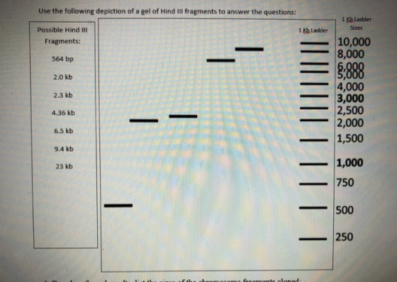
From the gel picture, we can find the approximate size of each fragment by matching them with those in the 1 Kb ladder.
1. There are 7 possible Hind III fragments listed out of which 5 are found in the gel. The corresponding sizes of the fragments in the gel are: 9.4 kb ,6.5 kb, 2.3 kb, 2.0 kb and 564 bp.
2. The fragment sizes that are not found in the list would be the remaining two listed, 4.36 kb and 23 Kb.
4. Based on the explanation in answer 3, it is evident that the two Hind III fragments that are not jagged should be the end fragments of the linear chromosome, meaning, these two fragments would be flanking the 5 fragments in the gel. But, in order to build a restriction map, it is necessary to have at least 2 enzymes cutting the DNA so that we are able to determine the relative postions of the fragments.
Add Answer to:
Use the following depiction of a gel of Hind III fragments to answer the questions: 1...
Based on the fragments cloned (and not cloned), draw the 45 kb hypothetical chromosome The Cloning...
 Based on the fragments cloned
(and not cloned), draw the 45 kb hypothetical chromosome
The Cloning Problem Week 9 Write-up Use the following depiction of a gel of Hind III fragments to answer the questions: 1 Kb Ladder Sizes 1 Kb Ladder Possible Hind III Fragments: 10,000 8,000 564 bp 8;888 2.0 kb 2.3 kb 4.36 kb 4,000 3,000 2,500 2,000 1,500 6.5 kb 9.4 kb |||||||||| 23 kb 1,000 750 S00 250
Based on the fragments cloned
(and not cloned), draw the 45 kb hypothetical chromosome
The Cloning Problem Week 9 Write-up Use the following depiction of a gel of Hind III fragments to answer the questions: 1 Kb Ladder Sizes 1 Kb Ladder Possible Hind III Fragments: 10,000 8,000 564 bp 8;888 2.0 kb 2.3 kb 4.36 kb 4,000 3,000 2,500 2,000 1,500 6.5 kb 9.4 kb |||||||||| 23 kb 1,000 750 S00 250
Genetics Yeast Lab The Awesome Power of Yeast Genetics Pre-lab questions Two weeks ago, you received...
 Genetics Yeast Lab
The Awesome Power of Yeast Genetics Pre-lab questions Two weeks ago, you received three unknown yeast strains. One was lys- (it cannot synthesize its own lysine) while the other two were LYS+. Because they were LYS+, they should be sensitive to high levels of aminoadipate (AA) before spontaneous and/or induced mutations occur that may allow some cells to become AA resistant. The two LYS+ strains differ in the rate at which they accumulate mutations. Two weeks ago,...
Genetics Yeast Lab
The Awesome Power of Yeast Genetics Pre-lab questions Two weeks ago, you received three unknown yeast strains. One was lys- (it cannot synthesize its own lysine) while the other two were LYS+. Because they were LYS+, they should be sensitive to high levels of aminoadipate (AA) before spontaneous and/or induced mutations occur that may allow some cells to become AA resistant. The two LYS+ strains differ in the rate at which they accumulate mutations. Two weeks ago,...
can someone explain throughly on how to find a-c??? thanks!!! The following question will provide practice in interpreting and analyzing gel results. 5. You obtained the DNA electrophoresis gel be...
 can
someone explain throughly on how to find a-c??? thanks!!!
The following question will provide practice in interpreting and analyzing gel results. 5. You obtained the DNA electrophoresis gel below. Three samples of lambda phage DNA were digested with 3 different restriction enzymes and the digested DNA was applied to the gel in lane 4 and the bands were visualized. The Hind Ill digest was used as a molecular weight standard marker and produced 6 DNA fragments of known size:...
can
someone explain throughly on how to find a-c??? thanks!!!
The following question will provide practice in interpreting and analyzing gel results. 5. You obtained the DNA electrophoresis gel below. Three samples of lambda phage DNA were digested with 3 different restriction enzymes and the digested DNA was applied to the gel in lane 4 and the bands were visualized. The Hind Ill digest was used as a molecular weight standard marker and produced 6 DNA fragments of known size:...
Hi I have a problem with number 5, it involves gel analysis results. There are 2 parts, a,b,c. For A Im sure you need to make a graph with distance in (cm) on the vertical axis and log10 bp on the hor...
 Hi I have a problem with number 5, it involves gel
analysis results. There are 2 parts, a,b,c. For A Im sure you need
to make a graph with distance in (cm) on the vertical axis and
log10 bp on the horitzontal. I need help figuring out where to
start and what to do. Please help!
The following question will provide practice in interpreting and analyzing gel results You obtained the DNA electrophoresis gel below. Three samples of lambda phage...
Hi I have a problem with number 5, it involves gel
analysis results. There are 2 parts, a,b,c. For A Im sure you need
to make a graph with distance in (cm) on the vertical axis and
log10 bp on the horitzontal. I need help figuring out where to
start and what to do. Please help!
The following question will provide practice in interpreting and analyzing gel results You obtained the DNA electrophoresis gel below. Three samples of lambda phage...
on the me but not with the larger fragments on o t and more and record...
 on the me but not with the larger fragments on o t and more and record med in the h 3. Determine the distracted for each bund data bere 4. Determine the distance traveled for each and generated in the double digest and record data bere tion Endonuclease Digestion and Gel Electrophoresis of DNA 185 VI. RESTRICTION MAPPING The different DNA fragments generated by different map DNA. For example, specific DNA on 2000 nucleotides in las Rl or Hind and...
colony which one express gene Only correct explanation not just answears pls. how to determine correct...
 colony which one express gene
Only correct explanation not just answears pls.
how to determine correct orientation by electrophoresis
CORI -Hindi mal kpn ! Aggi -Hind III Gene CDNA EcoRI 5. G' AAT TC 3 CTTAAG. 5 Бра 5GGT ACC 3 3 CCATGG 5 pMAE2 Avr 11 M5 CCTAGGS 3 GGATCC.5 Arell ACCGGT 3 3.. TGGCCA...5 Amp Hind 1 5: AAGCTT 3 S 3. TTCGAA53 Xma C CGGG.3 GGGCGC 5 You try the following strategies in order to see if...
colony which one express gene
Only correct explanation not just answears pls.
how to determine correct orientation by electrophoresis
CORI -Hindi mal kpn ! Aggi -Hind III Gene CDNA EcoRI 5. G' AAT TC 3 CTTAAG. 5 Бра 5GGT ACC 3 3 CCATGG 5 pMAE2 Avr 11 M5 CCTAGGS 3 GGATCC.5 Arell ACCGGT 3 3.. TGGCCA...5 Amp Hind 1 5: AAGCTT 3 S 3. TTCGAA53 Xma C CGGG.3 GGGCGC 5 You try the following strategies in order to see if...
Sketch the predicted migration pattern on an agarose gel of the following DNA samples: (1) Undigested...
Sketch the predicted migration pattern on an agarose gel of the following DNA samples: (1) Undigested pET15b plasmid; (2) pET15b digested with the EagI restriction enzyme; (3) pET15b digested with NdeI and AhdI (see pET15b vector map for location of these enzyme cutting sites). Include in your sketch a DNA base-pair ladder from 0 to 10,000 bp with 1000 bp increments. Label each band in the DNA ladder with the known size (in base-pairs), and estimate the size of the...
Hi can someone help me understand part C and why the drawn in red lines are...
 Hi can someone help me understand part C and why the
drawn in red lines are where they are.
Basically from the bp given how can I go back to cm so I can
drawn them into the picture provided?
Do not need help with A or B.
The following question will provide practice in interpreting and analyzing gel results You obtained the DNA electrophoresis gel below. Three samples of lambda phage DNA were digested with 3 different restriction enzymes...
Hi can someone help me understand part C and why the
drawn in red lines are where they are.
Basically from the bp given how can I go back to cm so I can
drawn them into the picture provided?
Do not need help with A or B.
The following question will provide practice in interpreting and analyzing gel results You obtained the DNA electrophoresis gel below. Three samples of lambda phage DNA were digested with 3 different restriction enzymes...
2 Addendum 1 to this lab provides information on the location of the EcoRI, and Sspl...
 2 Addendum 1 to this lab provides information on the location of the EcoRI, and Sspl cut sites in your BlueScript vector and the 1.3 kb EcoRI fragment. Using this information, (a) draw the two possible restriction maps for the recombinant pBlueKan plasmid that you constructed (see below). There are two possible maps because the 1.3 kb fragment could have been cloned in either orientation relative to the vector, Next, (b) predict the sizes of the EcoRI. and Sspl frements...
very lost on this whole worksheet and i dont know if my current answers are even...
 very lost on this whole worksheet and i dont know if my
current answers are even right, please help i need this done by
today
Name: Cloning Worksheet chromosome qut 756bp pBiochem 5.983 bp ladder Lane 1 Lane 2 Lane 3 Lane 4 Lane 5 Lane 6 BamHI (4233) (230) IT Enzyme recognition S ..GATATC. 51 EcoRV S .. . 3..CTATAC... 5 12 Pvull 89 BamHI Pett You are given the task to clone yfg into the plasmid pBioChem. Follow...
very lost on this whole worksheet and i dont know if my
current answers are even right, please help i need this done by
today
Name: Cloning Worksheet chromosome qut 756bp pBiochem 5.983 bp ladder Lane 1 Lane 2 Lane 3 Lane 4 Lane 5 Lane 6 BamHI (4233) (230) IT Enzyme recognition S ..GATATC. 51 EcoRV S .. . 3..CTATAC... 5 12 Pvull 89 BamHI Pett You are given the task to clone yfg into the plasmid pBioChem. Follow...
 Based on the fragments cloned
(and not cloned), draw the 45 kb hypothetical chromosome
The Cloning Problem Week 9 Write-up Use the following depiction of a gel of Hind III fragments to answer the questions: 1 Kb Ladder Sizes 1 Kb Ladder Possible Hind III Fragments: 10,000 8,000 564 bp 8;888 2.0 kb 2.3 kb 4.36 kb 4,000 3,000 2,500 2,000 1,500 6.5 kb 9.4 kb |||||||||| 23 kb 1,000 750 S00 250
Based on the fragments cloned
(and not cloned), draw the 45 kb hypothetical chromosome
The Cloning Problem Week 9 Write-up Use the following depiction of a gel of Hind III fragments to answer the questions: 1 Kb Ladder Sizes 1 Kb Ladder Possible Hind III Fragments: 10,000 8,000 564 bp 8;888 2.0 kb 2.3 kb 4.36 kb 4,000 3,000 2,500 2,000 1,500 6.5 kb 9.4 kb |||||||||| 23 kb 1,000 750 S00 250
 Genetics Yeast Lab
The Awesome Power of Yeast Genetics Pre-lab questions Two weeks ago, you received three unknown yeast strains. One was lys- (it cannot synthesize its own lysine) while the other two were LYS+. Because they were LYS+, they should be sensitive to high levels of aminoadipate (AA) before spontaneous and/or induced mutations occur that may allow some cells to become AA resistant. The two LYS+ strains differ in the rate at which they accumulate mutations. Two weeks ago,...
Genetics Yeast Lab
The Awesome Power of Yeast Genetics Pre-lab questions Two weeks ago, you received three unknown yeast strains. One was lys- (it cannot synthesize its own lysine) while the other two were LYS+. Because they were LYS+, they should be sensitive to high levels of aminoadipate (AA) before spontaneous and/or induced mutations occur that may allow some cells to become AA resistant. The two LYS+ strains differ in the rate at which they accumulate mutations. Two weeks ago,...
 can
someone explain throughly on how to find a-c??? thanks!!!
The following question will provide practice in interpreting and analyzing gel results. 5. You obtained the DNA electrophoresis gel below. Three samples of lambda phage DNA were digested with 3 different restriction enzymes and the digested DNA was applied to the gel in lane 4 and the bands were visualized. The Hind Ill digest was used as a molecular weight standard marker and produced 6 DNA fragments of known size:...
can
someone explain throughly on how to find a-c??? thanks!!!
The following question will provide practice in interpreting and analyzing gel results. 5. You obtained the DNA electrophoresis gel below. Three samples of lambda phage DNA were digested with 3 different restriction enzymes and the digested DNA was applied to the gel in lane 4 and the bands were visualized. The Hind Ill digest was used as a molecular weight standard marker and produced 6 DNA fragments of known size:...
 Hi I have a problem with number 5, it involves gel
analysis results. There are 2 parts, a,b,c. For A Im sure you need
to make a graph with distance in (cm) on the vertical axis and
log10 bp on the horitzontal. I need help figuring out where to
start and what to do. Please help!
The following question will provide practice in interpreting and analyzing gel results You obtained the DNA electrophoresis gel below. Three samples of lambda phage...
Hi I have a problem with number 5, it involves gel
analysis results. There are 2 parts, a,b,c. For A Im sure you need
to make a graph with distance in (cm) on the vertical axis and
log10 bp on the horitzontal. I need help figuring out where to
start and what to do. Please help!
The following question will provide practice in interpreting and analyzing gel results You obtained the DNA electrophoresis gel below. Three samples of lambda phage...
 on the me but not with the larger fragments on o t and more and record med in the h 3. Determine the distracted for each bund data bere 4. Determine the distance traveled for each and generated in the double digest and record data bere tion Endonuclease Digestion and Gel Electrophoresis of DNA 185 VI. RESTRICTION MAPPING The different DNA fragments generated by different map DNA. For example, specific DNA on 2000 nucleotides in las Rl or Hind and...
 colony which one express gene
Only correct explanation not just answears pls.
how to determine correct orientation by electrophoresis
CORI -Hindi mal kpn ! Aggi -Hind III Gene CDNA EcoRI 5. G' AAT TC 3 CTTAAG. 5 Бра 5GGT ACC 3 3 CCATGG 5 pMAE2 Avr 11 M5 CCTAGGS 3 GGATCC.5 Arell ACCGGT 3 3.. TGGCCA...5 Amp Hind 1 5: AAGCTT 3 S 3. TTCGAA53 Xma C CGGG.3 GGGCGC 5 You try the following strategies in order to see if...
colony which one express gene
Only correct explanation not just answears pls.
how to determine correct orientation by electrophoresis
CORI -Hindi mal kpn ! Aggi -Hind III Gene CDNA EcoRI 5. G' AAT TC 3 CTTAAG. 5 Бра 5GGT ACC 3 3 CCATGG 5 pMAE2 Avr 11 M5 CCTAGGS 3 GGATCC.5 Arell ACCGGT 3 3.. TGGCCA...5 Amp Hind 1 5: AAGCTT 3 S 3. TTCGAA53 Xma C CGGG.3 GGGCGC 5 You try the following strategies in order to see if...
 Hi can someone help me understand part C and why the
drawn in red lines are where they are.
Basically from the bp given how can I go back to cm so I can
drawn them into the picture provided?
Do not need help with A or B.
The following question will provide practice in interpreting and analyzing gel results You obtained the DNA electrophoresis gel below. Three samples of lambda phage DNA were digested with 3 different restriction enzymes...
Hi can someone help me understand part C and why the
drawn in red lines are where they are.
Basically from the bp given how can I go back to cm so I can
drawn them into the picture provided?
Do not need help with A or B.
The following question will provide practice in interpreting and analyzing gel results You obtained the DNA electrophoresis gel below. Three samples of lambda phage DNA were digested with 3 different restriction enzymes...
 2 Addendum 1 to this lab provides information on the location of the EcoRI, and Sspl cut sites in your BlueScript vector and the 1.3 kb EcoRI fragment. Using this information, (a) draw the two possible restriction maps for the recombinant pBlueKan plasmid that you constructed (see below). There are two possible maps because the 1.3 kb fragment could have been cloned in either orientation relative to the vector, Next, (b) predict the sizes of the EcoRI. and Sspl frements...
 very lost on this whole worksheet and i dont know if my
current answers are even right, please help i need this done by
today
Name: Cloning Worksheet chromosome qut 756bp pBiochem 5.983 bp ladder Lane 1 Lane 2 Lane 3 Lane 4 Lane 5 Lane 6 BamHI (4233) (230) IT Enzyme recognition S ..GATATC. 51 EcoRV S .. . 3..CTATAC... 5 12 Pvull 89 BamHI Pett You are given the task to clone yfg into the plasmid pBioChem. Follow...
very lost on this whole worksheet and i dont know if my
current answers are even right, please help i need this done by
today
Name: Cloning Worksheet chromosome qut 756bp pBiochem 5.983 bp ladder Lane 1 Lane 2 Lane 3 Lane 4 Lane 5 Lane 6 BamHI (4233) (230) IT Enzyme recognition S ..GATATC. 51 EcoRV S .. . 3..CTATAC... 5 12 Pvull 89 BamHI Pett You are given the task to clone yfg into the plasmid pBioChem. Follow...
Most questions answered within 3 hours.
-
Where is the error in this code sequence?
String s1 = "Hello";
String s2 = "ello";...
asked 11 months ago -
Financial data for Joel de Paris, Inc., for last year
follow:
Joel de Paris, Inc.
Balance...
asked 11 months ago -
Consider this reaction:
Al2(SO4)3 (aq)+ BaCl3
(aq) Al2Cl6 (aq)- +
3BaSO4(s) . What is the...
asked 11 months ago -
Suppose that Savneet is considering increasing her
recent random sample from 20 car rentals to 40...
asked 11 months ago -
Trucks arrive at an unloading terminal at an average rate of 120
per hour.
Trucks arrive...
asked 11 months ago -
Why are methanol and ethanol completely soluble in water while
octanol is not very little soluble....
asked 11 months ago -
A facilities manager at a university reads in a research report
that the mean amount of...
asked 11 months ago -
When the CuSO4 is rehydrated by adding water to the anhydrous
compound, is this an endothermic...
asked 11 months ago -
A ray of sunlight is passing from diamond into crown glass; the
angle of incidence is...
asked 11 months ago -
A block of mass 0.249 kg is placed on top of a light, vertical
spring of...
asked 11 months ago -
how do the kidneys compensate in the presences of acidosis
a) trigger hyperventilate
b) reserve acid...
asked 11 months ago -
Question 501 pts
The rental rate of capital to the firm increases. Which of the
following...
asked 11 months ago