If you had to analyze bands on an agarose gel, can you differentiate a homozygous knockout,...
If you had to analyze bands on an agarose gel, can you differentiate a homozygous knockout, homozygous wild type and heterozygous genotype if you are given the different sized PCR fragments for knockout and wild type fragments?
Homework Answers
To solve this we have to keep in mind that to determine gene knockout, we have to identify a section of our gene representing the wild type and the same should be absent in the knockout condition in the PCR product and this PCR product should be generated by the same pair of primers as that of others. Another way to find out the absence in KO condition by looking for a shorter PCR product in the KO compared to wildtype.
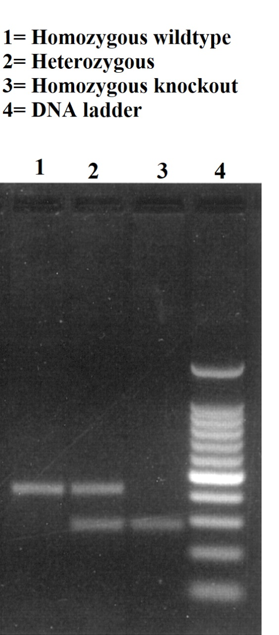
For wildtype homozygous condition we will get a single PCR product i.e., we will see one strong band of the size of our wild type PCR product.
For KO homozygous condition, we will get one strong band generally smaller than wild type (in the gel it appears to be present below homozygous wild type).
For heterozygous condition, we will get two bands: one at the wild type size and one at the KO size.
Add Answer to:
If you had to analyze bands on an agarose gel, can you
differentiate a homozygous knockout,...
The picture above represents an agarose gel that was used to analyze plasmid DNA after it...
 The picture above represents an agarose gel that was used to analyze plasmid DNA after it was cut with the restriction enzyme HindIll. The plasmid was incubated with Hindill until all of the available Hindlll cut sites were cut by HindIll. After running the sample on the gel, three bands were detected (Note that there are three wells shown at the top of the gel for loading samples, however, only the middle well was loaded with sample). Based on this...
B. To examine whether the PCR was successful, you ran an agarose gel. The expected length of your...
 Solution please
B. To examine whether the PCR was successful, you ran an agarose gel. The expected length of your targeted gene was 1,200 base pairs (BP) Lane 10,000 BP | - 4,000 BP 2,000 BP 800 BP : 400 BP A C 1. What were bands A, B, and C? (1 pt)
B. To examine whether the PCR was successful, you ran an agarose gel. The expected length of your targeted gene was 1,200 base pairs (BP) Lane 10,000...
Solution please
B. To examine whether the PCR was successful, you ran an agarose gel. The expected length of your targeted gene was 1,200 base pairs (BP) Lane 10,000 BP | - 4,000 BP 2,000 BP 800 BP : 400 BP A C 1. What were bands A, B, and C? (1 pt)
B. To examine whether the PCR was successful, you ran an agarose gel. The expected length of your targeted gene was 1,200 base pairs (BP) Lane 10,000...
When looking at the results of the negative control on the agarose gel, what do you...
When looking at the results of the negative control on the agarose gel, what do you expect to see? Select one: a. The products will be the same size as the blue and brown eye colour samples but the bands visible will be more faint (lighter). b. You will see fewer bands when compared to the blue and brown eye colour samples. c. There should be no PCR product visible. d. You will see a PCR product, but it will...
Can anyone show me step-by-step on how to do the agarose calculations? This week we will...
 Can
anyone show me step-by-step on how to do the agarose calculations?
This week we will run the PCR reactions on an agarose gel and analyze the results. Safety notes: Ethidium bromide is a potent mutagen. Wear gloves when handling containers containing ethidium bromide, when handling gels that have been stained in ethidium bromide, and when working with the computer attached to the gel-doc system. Protocol I. Pouring agarose gels I. Using 10x TAE, prepare 50 ml of 3% (w/v)...
Can
anyone show me step-by-step on how to do the agarose calculations?
This week we will run the PCR reactions on an agarose gel and analyze the results. Safety notes: Ethidium bromide is a potent mutagen. Wear gloves when handling containers containing ethidium bromide, when handling gels that have been stained in ethidium bromide, and when working with the computer attached to the gel-doc system. Protocol I. Pouring agarose gels I. Using 10x TAE, prepare 50 ml of 3% (w/v)...
Chromosomal and plasmid DNA can be cut into manageable pieces by restriction enzymes. Using agarose gel...
 Chromosomal and plasmid DNA can be cut into manageable pieces by
restriction enzymes. Using agarose gel electrophoresis, the DNA
fragments can be separated on a gel, based on their lengths. In
order to see the fragments, a stain is typically added to the gel.
The size of each fragment can be determined by comparing each one
to a DNA molecular weight marker of known size.
Below is a map of pBR22 plasmid. The position and base pair
number of the...
Chromosomal and plasmid DNA can be cut into manageable pieces by
restriction enzymes. Using agarose gel electrophoresis, the DNA
fragments can be separated on a gel, based on their lengths. In
order to see the fragments, a stain is typically added to the gel.
The size of each fragment can be determined by comparing each one
to a DNA molecular weight marker of known size.
Below is a map of pBR22 plasmid. The position and base pair
number of the...
Using Agarose Gel Electrophoresis to identify Hemoglobin Phenotypes 13. Draw the banding patterns you obtained on...
 Using Agarose Gel Electrophoresis to identify Hemoglobin Phenotypes 13. Draw the banding patterns you obtained on the space below. Sample 1. A Banding pattern Well 1.0 lbo 2. AS 3. S 4. Unknown 6. O 7. 5 7.0 8. Unknown 8. O 14. Why does sickle cell hemoglobin behave differently from normal hemoglobin during agarose gel electrophoresis? 15. + A child is born with sickle cell disease. What can you conclude about the potential genotypes of the parents? 16. +...
how can you tell the difference between a homozygous individual and a heterozygous individual for a...
how can you tell the difference between a homozygous individual and a heterozygous individual for a particular bi-allegic gene when looking at the results of a gel
can someone explain throughly on how to find a-c??? thanks!!! The following question will provide practice in interpreting and analyzing gel results. 5. You obtained the DNA electrophoresis gel be...
 can
someone explain throughly on how to find a-c??? thanks!!!
The following question will provide practice in interpreting and analyzing gel results. 5. You obtained the DNA electrophoresis gel below. Three samples of lambda phage DNA were digested with 3 different restriction enzymes and the digested DNA was applied to the gel in lane 4 and the bands were visualized. The Hind Ill digest was used as a molecular weight standard marker and produced 6 DNA fragments of known size:...
can
someone explain throughly on how to find a-c??? thanks!!!
The following question will provide practice in interpreting and analyzing gel results. 5. You obtained the DNA electrophoresis gel below. Three samples of lambda phage DNA were digested with 3 different restriction enzymes and the digested DNA was applied to the gel in lane 4 and the bands were visualized. The Hind Ill digest was used as a molecular weight standard marker and produced 6 DNA fragments of known size:...
3) Image from Biotechnology Explorer Kit, BIO RAD Analysis of DNA Fragments The data you entered...
 3) Image from Biotechnology Explorer Kit, BIO RAD Analysis of DNA Fragments The data you entered for the lambda Hindill digest were the relative positions of DNA bands of known size. Since the exact size and position of these fragments are known, they can be used as standard reference points to estimate the size of unknown fragment bands. A set of fragments of known sizes is called a molecular weight ruler or standards or marker (or sometimes a ladder because...
Can you please help me with this problem: 3. A dominant wild-type allele D produces full...
 Can you please help me with this problem:
3. A dominant wild-type allele D produces full enzyme function, but a recessive alleled, produces no functional enzymatic action, and a recessive allele da produces reduced enzyme function. Western blot analysis of the proteins produced by organisms with different genotypes for this gene gives the results shown. Note, proteins separate out in a gel by size similar to DNA fragments with smaller polypeptides traveling farther. Genotype DD Dd, Dd, dod, dd₂ d₂d₂...
Can you please help me with this problem:
3. A dominant wild-type allele D produces full enzyme function, but a recessive alleled, produces no functional enzymatic action, and a recessive allele da produces reduced enzyme function. Western blot analysis of the proteins produced by organisms with different genotypes for this gene gives the results shown. Note, proteins separate out in a gel by size similar to DNA fragments with smaller polypeptides traveling farther. Genotype DD Dd, Dd, dod, dd₂ d₂d₂...
 The picture above represents an agarose gel that was used to analyze plasmid DNA after it was cut with the restriction enzyme HindIll. The plasmid was incubated with Hindill until all of the available Hindlll cut sites were cut by HindIll. After running the sample on the gel, three bands were detected (Note that there are three wells shown at the top of the gel for loading samples, however, only the middle well was loaded with sample). Based on this...
 Solution please
B. To examine whether the PCR was successful, you ran an agarose gel. The expected length of your targeted gene was 1,200 base pairs (BP) Lane 10,000 BP | - 4,000 BP 2,000 BP 800 BP : 400 BP A C 1. What were bands A, B, and C? (1 pt)
B. To examine whether the PCR was successful, you ran an agarose gel. The expected length of your targeted gene was 1,200 base pairs (BP) Lane 10,000...
Solution please
B. To examine whether the PCR was successful, you ran an agarose gel. The expected length of your targeted gene was 1,200 base pairs (BP) Lane 10,000 BP | - 4,000 BP 2,000 BP 800 BP : 400 BP A C 1. What were bands A, B, and C? (1 pt)
B. To examine whether the PCR was successful, you ran an agarose gel. The expected length of your targeted gene was 1,200 base pairs (BP) Lane 10,000...
 Can
anyone show me step-by-step on how to do the agarose calculations?
This week we will run the PCR reactions on an agarose gel and analyze the results. Safety notes: Ethidium bromide is a potent mutagen. Wear gloves when handling containers containing ethidium bromide, when handling gels that have been stained in ethidium bromide, and when working with the computer attached to the gel-doc system. Protocol I. Pouring agarose gels I. Using 10x TAE, prepare 50 ml of 3% (w/v)...
Can
anyone show me step-by-step on how to do the agarose calculations?
This week we will run the PCR reactions on an agarose gel and analyze the results. Safety notes: Ethidium bromide is a potent mutagen. Wear gloves when handling containers containing ethidium bromide, when handling gels that have been stained in ethidium bromide, and when working with the computer attached to the gel-doc system. Protocol I. Pouring agarose gels I. Using 10x TAE, prepare 50 ml of 3% (w/v)...
 Chromosomal and plasmid DNA can be cut into manageable pieces by
restriction enzymes. Using agarose gel electrophoresis, the DNA
fragments can be separated on a gel, based on their lengths. In
order to see the fragments, a stain is typically added to the gel.
The size of each fragment can be determined by comparing each one
to a DNA molecular weight marker of known size.
Below is a map of pBR22 plasmid. The position and base pair
number of the...
Chromosomal and plasmid DNA can be cut into manageable pieces by
restriction enzymes. Using agarose gel electrophoresis, the DNA
fragments can be separated on a gel, based on their lengths. In
order to see the fragments, a stain is typically added to the gel.
The size of each fragment can be determined by comparing each one
to a DNA molecular weight marker of known size.
Below is a map of pBR22 plasmid. The position and base pair
number of the...
 Using Agarose Gel Electrophoresis to identify Hemoglobin Phenotypes 13. Draw the banding patterns you obtained on the space below. Sample 1. A Banding pattern Well 1.0 lbo 2. AS 3. S 4. Unknown 6. O 7. 5 7.0 8. Unknown 8. O 14. Why does sickle cell hemoglobin behave differently from normal hemoglobin during agarose gel electrophoresis? 15. + A child is born with sickle cell disease. What can you conclude about the potential genotypes of the parents? 16. +...
 can
someone explain throughly on how to find a-c??? thanks!!!
The following question will provide practice in interpreting and analyzing gel results. 5. You obtained the DNA electrophoresis gel below. Three samples of lambda phage DNA were digested with 3 different restriction enzymes and the digested DNA was applied to the gel in lane 4 and the bands were visualized. The Hind Ill digest was used as a molecular weight standard marker and produced 6 DNA fragments of known size:...
can
someone explain throughly on how to find a-c??? thanks!!!
The following question will provide practice in interpreting and analyzing gel results. 5. You obtained the DNA electrophoresis gel below. Three samples of lambda phage DNA were digested with 3 different restriction enzymes and the digested DNA was applied to the gel in lane 4 and the bands were visualized. The Hind Ill digest was used as a molecular weight standard marker and produced 6 DNA fragments of known size:...
 3) Image from Biotechnology Explorer Kit, BIO RAD Analysis of DNA Fragments The data you entered for the lambda Hindill digest were the relative positions of DNA bands of known size. Since the exact size and position of these fragments are known, they can be used as standard reference points to estimate the size of unknown fragment bands. A set of fragments of known sizes is called a molecular weight ruler or standards or marker (or sometimes a ladder because...
 Can you please help me with this problem:
3. A dominant wild-type allele D produces full enzyme function, but a recessive alleled, produces no functional enzymatic action, and a recessive allele da produces reduced enzyme function. Western blot analysis of the proteins produced by organisms with different genotypes for this gene gives the results shown. Note, proteins separate out in a gel by size similar to DNA fragments with smaller polypeptides traveling farther. Genotype DD Dd, Dd, dod, dd₂ d₂d₂...
Can you please help me with this problem:
3. A dominant wild-type allele D produces full enzyme function, but a recessive alleled, produces no functional enzymatic action, and a recessive allele da produces reduced enzyme function. Western blot analysis of the proteins produced by organisms with different genotypes for this gene gives the results shown. Note, proteins separate out in a gel by size similar to DNA fragments with smaller polypeptides traveling farther. Genotype DD Dd, Dd, dod, dd₂ d₂d₂...
Most questions answered within 3 hours.
-
Where is the error in this code sequence?
String s1 = "Hello";
String s2 = "ello";...
asked 11 months ago -
Financial data for Joel de Paris, Inc., for last year
follow:
Joel de Paris, Inc.
Balance...
asked 11 months ago -
Consider this reaction:
Al2(SO4)3 (aq)+ BaCl3
(aq) Al2Cl6 (aq)- +
3BaSO4(s) . What is the...
asked 11 months ago -
Suppose that Savneet is considering increasing her
recent random sample from 20 car rentals to 40...
asked 11 months ago -
Trucks arrive at an unloading terminal at an average rate of 120
per hour.
Trucks arrive...
asked 11 months ago -
Why are methanol and ethanol completely soluble in water while
octanol is not very little soluble....
asked 11 months ago -
A facilities manager at a university reads in a research report
that the mean amount of...
asked 11 months ago -
When the CuSO4 is rehydrated by adding water to the anhydrous
compound, is this an endothermic...
asked 11 months ago -
A ray of sunlight is passing from diamond into crown glass; the
angle of incidence is...
asked 11 months ago -
A block of mass 0.249 kg is placed on top of a light, vertical
spring of...
asked 11 months ago -
how do the kidneys compensate in the presences of acidosis
a) trigger hyperventilate
b) reserve acid...
asked 11 months ago -
Question 501 pts
The rental rate of capital to the firm increases. Which of the
following...
asked 11 months ago